


Influential Discoveries
As told by Winfried Denk
My father grew up on a farm near Brno, in a part of Czechoslowakia where a lot of ethnic Germans were then living. After the war he could not return there, became a refugee, and was placed to work and live on a farm near the town of Kempten, where he met my mother. They got married and moved to Ottobrunn, building the house I was to grow up in. I was born in Munich, in November 1957.

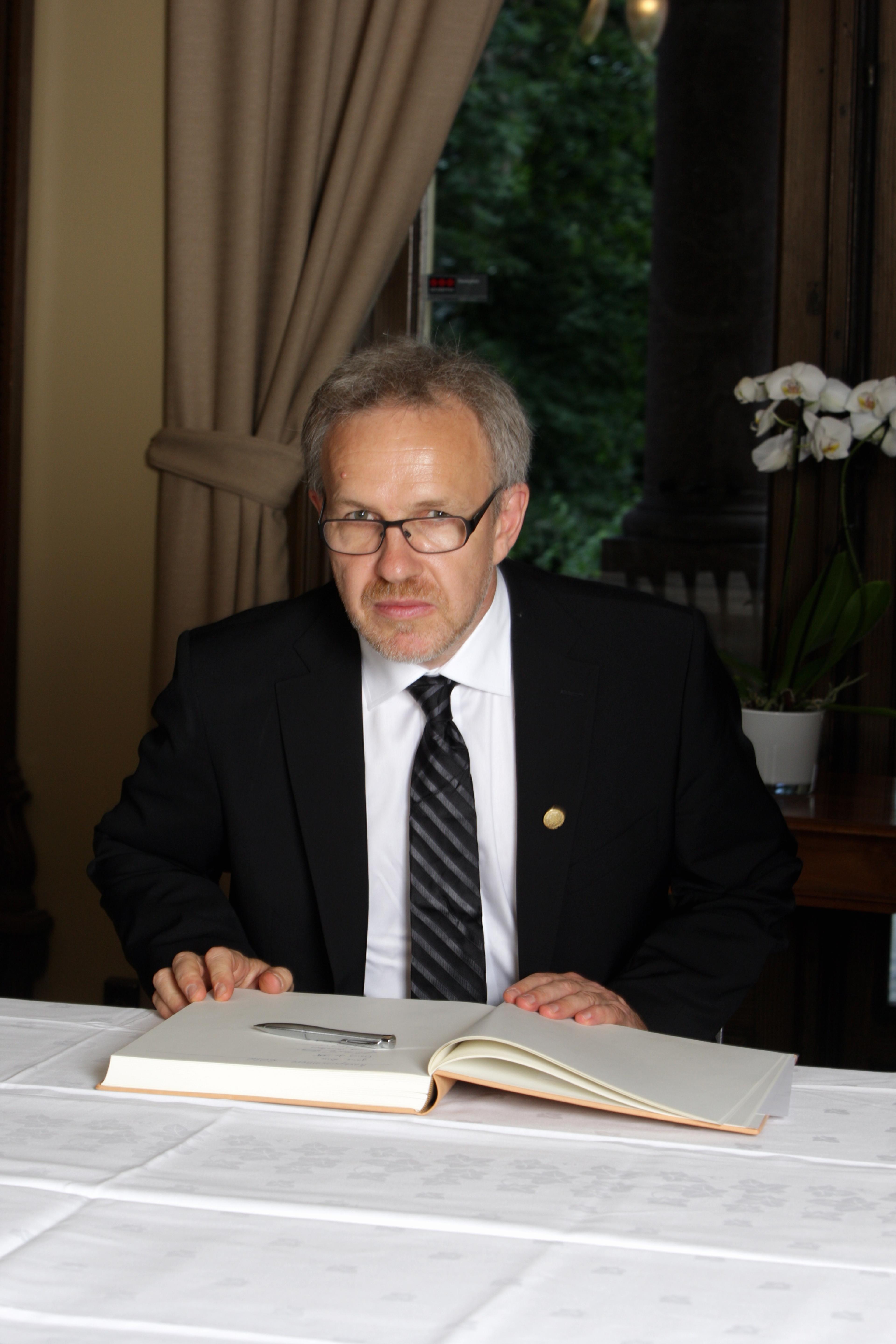
Winfried Denk signing the guest book at the Norwegian Academy of Science and Letters during the Kavli Prize week in Oslo (Photo credit: Eirik Furu Baardsen).
My father set up a workshop in the basement, outfitted for wood lathing, a craft he had studied. As a child I spent much time in this workshop, learning how use tools and build things. My parents appreciated education and strongly encouraged my younger sister and me to get as much of it as possible. After finishing elementary school I entered the Asam Gymnasium in Munich, where it became apparent that my talents were unevenly spread across subjects, math and physics being favored. I was an unremarkable student and spent a lot of time on extracurricular activities. I fixed and built electronic devices, got involved in the youth organization of the conservative party (an early act of rebellion amid my mostly left-leaning peers) and edited a student newspaper. A few great teachers, among them Mr. Lanz in physics and Mr. Hilgarth in biology, kept my interest in science alive. A big influence was a close friend’s father, a Professor for Electrical Engineering, who instilled in me a passion for electronics and was an early academic role model. Unsure for a while, in the end I decided to become a physicist rather than electrical engineer, choosing the more fundamental subject. In 1978, after the mandatory 15 months in the army, I enrolled in the Ludwigs-Maximilians University in Munich. Again, much time was spent on extracurricular activities, mostly on preparations for a trip to the Sahara desert, which involved retrofitting two surplus army trucks with diesel engines.
IBM research lab
In 1981 I moved to the ETH in Zurich, vaguely inspired by the fact that Einstein had studied there but mostly driven by the desire to finally get away from home. This being Switzerland, I needed to earn some extra money and was very lucky to find a job as an undergraduate researcher at the IBM research lab in Rüschlikon, a suburb of Zurich. I had never been particularly motivated to spend much time on problem sets or lab exercises; getting to do original research in a lab finally focused my efforts on science. The IBM research lab was where, at that time, the Scanning Tunneling Microscope was being developed by Gerd Binning and his colleagues. Dieter Pohl, whom I worked for, had the idea to build a super-resolution optical microscope by scanning a sub-wavelength aperture along a surface. One problem was how to make a small, nanometer sized hole. An idea I had, to push an aluminum-covered quartz tip against a glass slide until light started coming through, worked, allowing us to produce the first super-resolution optical images. It was probably then that I developed a passion for scanning microscopy. In order to graduate I needed to write a Master’s thesis, which I did in Kurth Wüthrich’s lab under the direct guidance of Gerhard Wagner. I liked structural biology and learned a lot about biological macromolecules but I also felt that NMR spectroscopy was not my thing because it didn’t involve enough building of experimental apparatus.

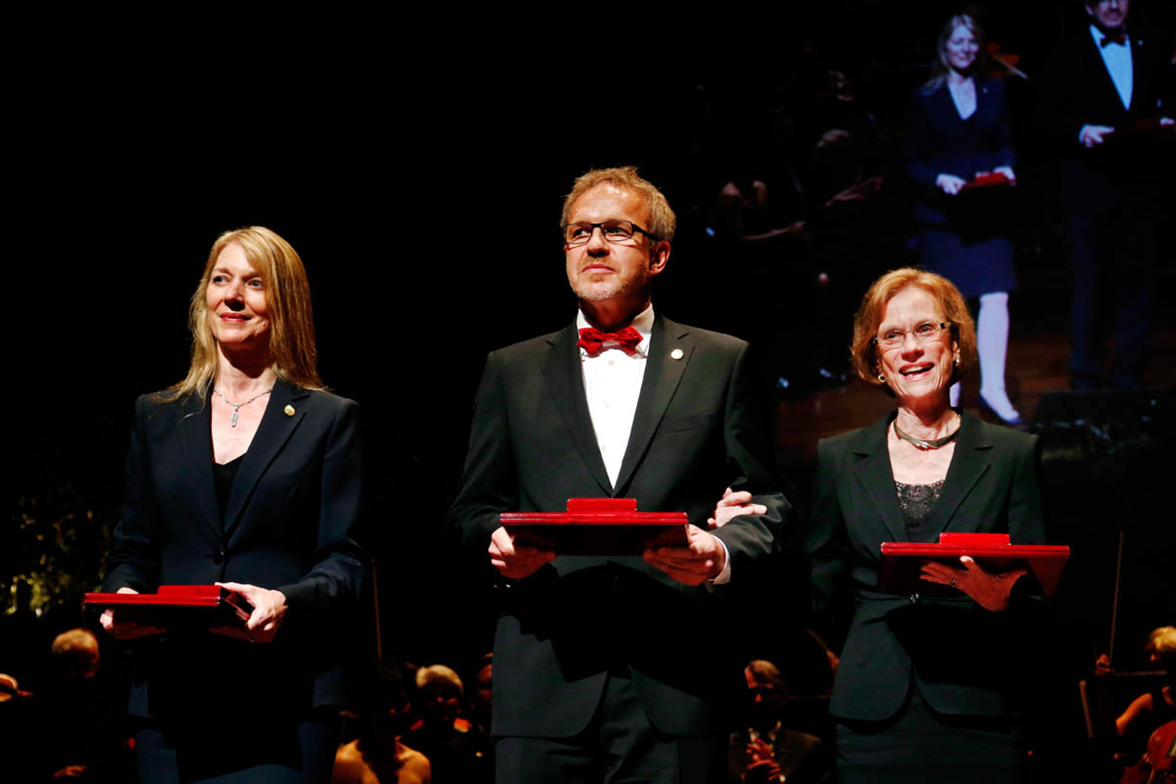
2012 Kavli Prize Neuroscience laureates at the ceremony in Oslo, from left: Cornelia Bargmann, Winfried Denk and Ann Martin Graybiel (Photo credit: Scanpix).
Traveling across the Atlantic
Next, I wanted to go to the United States, then and arguably still the Mecca of science. Berkeley, my first choice, did not accept me (fortunately, in retrospect) and I had to go to Cornell, my backup school, where I joined Watt Webb’s lab. Webb had an unusual academic background; he had worked at Union Carbide before coming to Cornell and had been a material scientist before turning to biophysics. His lab was very interested in methods – both fluorescence-correlation and photo bleaching-recovery spectroscopy had been invented there – and he gave students and postdocs a lot of freedom. Webb first wanted me to work on voltage-sensitive dyes, which didn’t particularly capture my interest, and he almost fired me after I spent 6 weeks in Greece, studying monk seals. Giving me a second chance, he suggested I do a project that involved measuring the motion of sensory hair bundles in the inner ear. This was interesting, mainly because I would be spending some time in San Francisco, ostensibly to learn how to prepare the tissue in Jim Hudspeth’s lab. From Hudspeth I also learned how to do physiology well and how to answer a question definitively. Back in Ithaca I began doing experiments on hair cells, ultimately showing that their extreme sensitivity enables them to hear their own Browning motion. Unrelated to my thesis work but important for things to come, Webb’s lab taught me all about the importance of fluorescence imaging, the problem of photo bleaching and photo damage, and that one could “image” calcium, using fluorescent indicators.
On one of my forays into the campus book store, I stumbled upon “Theory and Practice of Scanning Optical Microscopy” by Wilson and Sheppard. I bought it and read it cover to cover. This was around 1986, when commercial confocal microscopes were just becoming available but none existed in the building where I worked and probably on the entire campus. All work in the lab was done using widefield microscopy. As a result of my reading Wilson and Sheppard and talking about it, Webb bought a Biorad MRC 500 laser scanning microscope. One problem was that it would not work with the calcium indicators then available, which needed ultraviolet light for excitation. The book also talked about nonlinear optical effects, mostly second harmonic microscopy, but mentioned two-photon absorption only in passing. Two-photon excitation would not only solve the problem of exciting calcium indicators, but also give us optical sectioning while reducing photo damage away from the focal plane. Doing some back-ofthe-envelope calculations convinced me that enough fluorescence could be generated for imaging if one used a mode-locked laser. But I had a thesis to finish and so Jim Strickler, a new graduate student in the lab, got handed the project. I followed Jim’s progress: he did some background research and discovered that there was now a colliding-pulse mode-locked dye laser in the building, in the lab of a new faculty member, Frank Wise. So when I had to cool my heels after handing in my thesis, in May 1989, and before heading back to Switzerland in the fall, I suggested that we change some filters and beam splitters in the MRC 500s and take it up to Wise’s lab to see whether we could get some two-photon images. Alas, it worked right away and a few days later we had acquired almost all the data for the 1990 Science paper.
I went back to the IBM lab because I wanted to return to Europe and because I had liked working with Dieter Pohl as a student. As a postdoc I tried to combine near-field imaging with two-photon excitation, made some calculations but did not succeed in taking any images. I did find a way to measure local electrical conductivity by detecting the damping in a force microscope. Cured of my nostalgia for Europe and enticed by an offer from the famed Bell Laboratories to start my own lab, I moved to New Jersey early in 1991. My main concern was that Bell Labs might not be an ideal place to find a girlfriend. But it turned out to be the place where, in 1992, I met Renate Lobnig, who would become my wife, and our daughters, Marlis and Helena, were born in Newark in 1993 and 1995. At Bell I was surrounded by a small group of outstanding neuroscientists led by David Tank. Our lunch-table conversations were my neuroscience school. Returning to 2-photon excitation, I remembered that it could drive photochemical reactions, making it possible to map the distribution of neurotransmitter receptors by uncaging. Painful at that time because the caged compounds were too insensitive, two-photon scanning photochemical microscopy has now, with better compounds, found its niche.

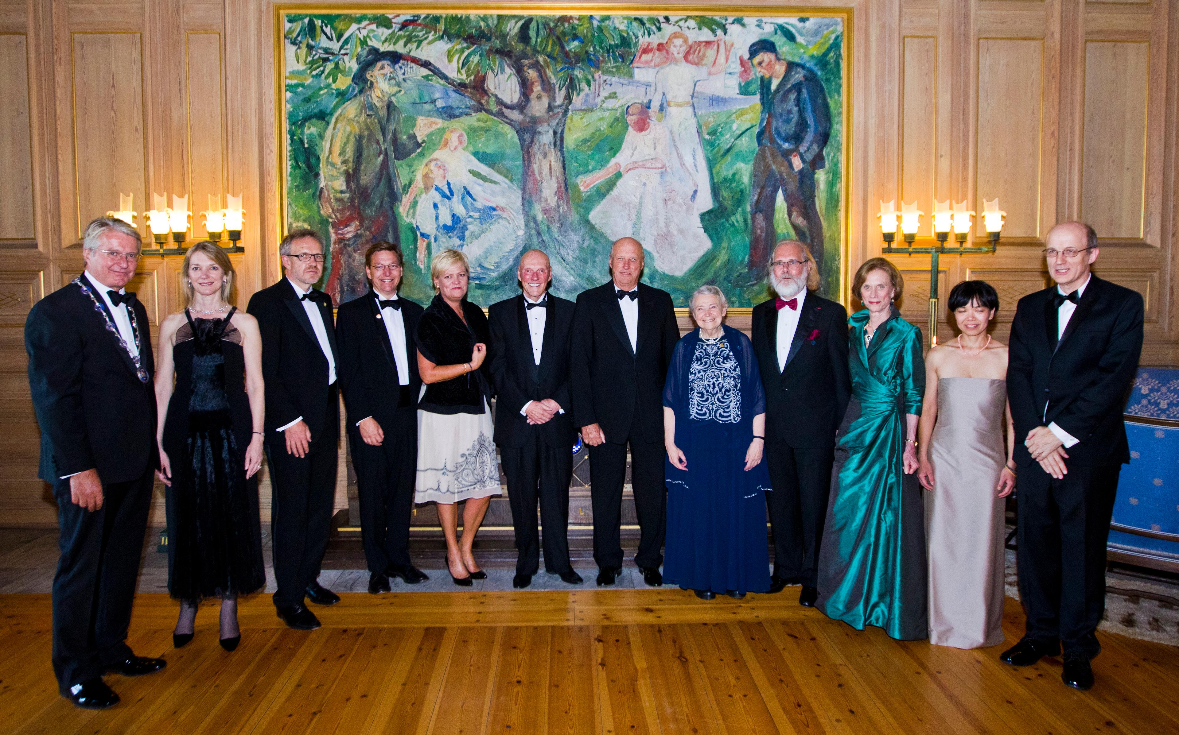
Reception in the Munch room at Oslo City Hall, from left: Mayor of Oslo Fabian Stang, Cornelia Bargmann (Kavli Prize neuroscience laureate), Winfried Denk (Kavli Prize neuroscience laureate), Michael Brown (Kavli Prize astrophysics laureate), Kristin Halvorsen (Norwegian minister of science and education), Fred Kavli, His Royal Highness King Harald, Mildred S. Dresselhaus (Kavli Prize nanoscience laureate), Nils Christian Stenseth (President of the Norwegian Academy of Science and Letters), Ann Martin Graybiel (Kavli Prize neuroscience laureate), Jane X. Luu (Kavli Prize astrophysics laureate) and David Jewitt (Kavli Prize astrophysics laureate). (Photo credit: Scanpix).
The two-photon microscopy
Soon the mode-locked dye laser was replaced by the titanium-sapphire laser, which was much easier to use and had longer wavelength, both essential for two-photon microscopy to become widely used. The TiSa’s wavelength range allowed 2-photon excitation of most common fluorophores, including GFP and the improved “green” calcium indicators. The key insight in those years was that, because it does not need a detector pinhole for optical sectioning, two-photon microscopy has a huge sensitivity advantage over confocal microscopy in scattering tissue. Rafael Yuste, now a professor at Columbia University, then a postdoc in Tank’s lab, across the corridor from mine, worked on brain slices and had (and still has) a passion for dendritic spines. We took movies of the calcium dynamics in individual spines, which convinced many that two-photon microscopy was more than a gimmick. In 1993 or so, Karel Svoboda (now a group leader at Janelia Farm) joined the biological-computation department, as the neuroscience group was called, as a postdoc. We had crossed paths before, when he used an interferometer design that I developed to measure hair-bundle motion to watch motor molecules step. First, Karel measured diffusion in and out of spines using two-photon uncaging and photobleaching. Then he did the lion’s share of experiments that demonstrated two-photon imaging in the living brain, a project that also involved David Kleinfeld and David Tank. Later, another postdoc, Fritjof Helmchen (now at the University of Zurich), extended in vivo 2-photon imaging to freely moving rats. I still remember Bell Labs as a scientific paradise. We all worked closely together in varying constellations.
In 1997 Peter Detwiler, a physiologist from the University of Washington in Seattle, came to Bell Labs for a sabbatical. Right away we started working on the retina, getting nowhere at first. However, after switching from the goldfish to the salamander, we eventually managed to show that with the two-photon microscope one can image calcium signals without blinding the retina, which meant we could record responses to visual stimuli. This ended up getting me interested in the retina̶–not a part of the nervous system highly respected by cortico-supremacists–more deeply than I ever intended to.
The Max-Planck Institutes
Growing up in Germany, I was very much in awe of the Max-Planck Institutes; while in high school, I had visited the Martinried campus during one of the days when it was open to the public. So I was thrilled when in 1999, while I was in Berkeley serving on a review panel, Bert Sakmann called to tell me that I would be receiving an offer to become a Director at the Max-Planck Institute for Medical Research in Heidelberg. It was very fortunate that I could entice Rainer Friedrich (now at the Friedrich-Miescher Institute in Basel) to establish an independent junior group associated with the department. I was also very lucky to be joined by Thomas Euler (now a professor in Tübingen), a true retina expert. This allowed me to continue the retina work started at Bell Labs. Thomas then famously showed that direction selectivity is present in starburst amacrine-cell dendrites.
With some excellent physics graduate students from the University of Heidelberg, I continued to develop two-photon imaging technology; Patrick Theer was working on depth-penetration improvement, and Marcus Feierabend and Markus Rueckel worked on adaptive optics-based correction of tissue-induced distortion of the laser focus. But I felt that in the end those were more incremental advances and that the application of two-photon microscopy was now limited more by tissue preparation and staining technology, areas where I lacked talent. I was also frustrated by the fact that it is hard to draw definite conclusions from activity measurements about how a particular computation is actually performed. From my days of fixing broken electronics as a teenager I knew how useful circuit diagrams could be, and I was aware of Sidney Brenner’s reconstruction of the complete circuit diagram of “the worm”.
Connectomics
One day, Bert Sakmann (who had gotten into anatomical reconstructions of the neurons his lab had recorded from) challenged me to come up with a light-microscope design that would allow him to definitively identify synaptic contacts. I told him that the wavelength is just too long and that it couldn’t be done. But then I remembered a lunch table conversation at Bell Labs, instigated by David Tank, where we had discussed using scanning near-field microscopy (then being perfected by Eric Betzig at Bell Labs) to image the surface of a block of plastic-embedded tissue and then repeating the process after cutting a thin slice off the top and so forth. The prospect of using a technique as finicky and slow as probescanning microscopy did not appeal to me but I realized that one could use a scanning electron microscope instead. What was needed was to get a microtome into the chamber of the SEM. Fortunately Bert Sakmann allowed Heinz Horstmann, an enterprising and enthusiastic EM technician in his department, to spend time teaching me EM preparation techniques, adding to and refreshing the knowledge from a course on electron microscopy I had taken, on a whim, back at Cornell. Taking advantage of some recently acquired AutoCad skills, I designed an ultramicrotome to go into the SEM and had it built by the outstanding mechanical workshop at the institute. As it turned out, Steve Leighton had built such a microtome at Woods Hole several decades earlier, but the SEMs in those days were not good enough to provide the resolution and signal quality needed for circuit reconstruction and there was no way to deal with the amount of data that would be generated.
Again I was very lucky; two exceptional young scientists joined the Lab to work with me on what has since become known as connectomics. Moritz Helmstaeder, who is now a group leader at the Max-Planck Institute for Neurobiology in Martinsried and trained as both a medical doctor and a physicist, took on the task of analyzing the vast amount of data that was going to be generated by the Serial Block-Face Scanning Electron Microscope. Kevin Briggman (now at the NIH), a neuroscientist and an engineer, started optimizing the microtome and continued the development of the special staining techniques started by Heinz Horstmann. A biological question was found, surprise, surprise, in the direction selectivity circuit of the retina. What’s left in the retina is to finish a connectome.
Further ambitions
The ambition for the next decade (assuming my Parkinson’s disease keeps progressing slowly) is to get the data that allows reconstruction of the circuit diagram for a whole mouse brain. I am also looking forward to moving my department to Martinsried, probably in early 2015.
As early as 2004 I got involved in the planning process for the Howard Hughes Medical Institute’s Janelia Farm campus in northern Virginia. Now in operation, Janelia, partly inspired by Bell Labs, is committed to promoting an atmosphere of vigorous intellectual exchange and the development of new methods. For me it has become a second scientific home.