


Student Interactions Still Inspire Me
As told by Robert Fettiplace
The Fettiplace surname is reputed to be derived from ‘faîtes place’, an old French term for an usher. History indicates my ancestors accompanied William the Conqueror during the French invasion of England in 1066, and they were subsequently rewarded with much land in Oxfordshire and Berkshire, one becoming mayor of the city of Oxford in 1245. They are commemorated in trios of striking stone effigies of reclining Tudor and Stuart knights in St. Mary’s Church in Swinbrook, near Oxford. However, the dynasty fell on hard times in the 1700’s, and the family manor was occupied by highwaymen and subsequently burned to the ground. The last of the Oxford Fettiplaces expired in 1805, but a line had earlier escaped north and flourished in Nottingham, where I was born in 1946 to Bob and Maisie Fettiplace.

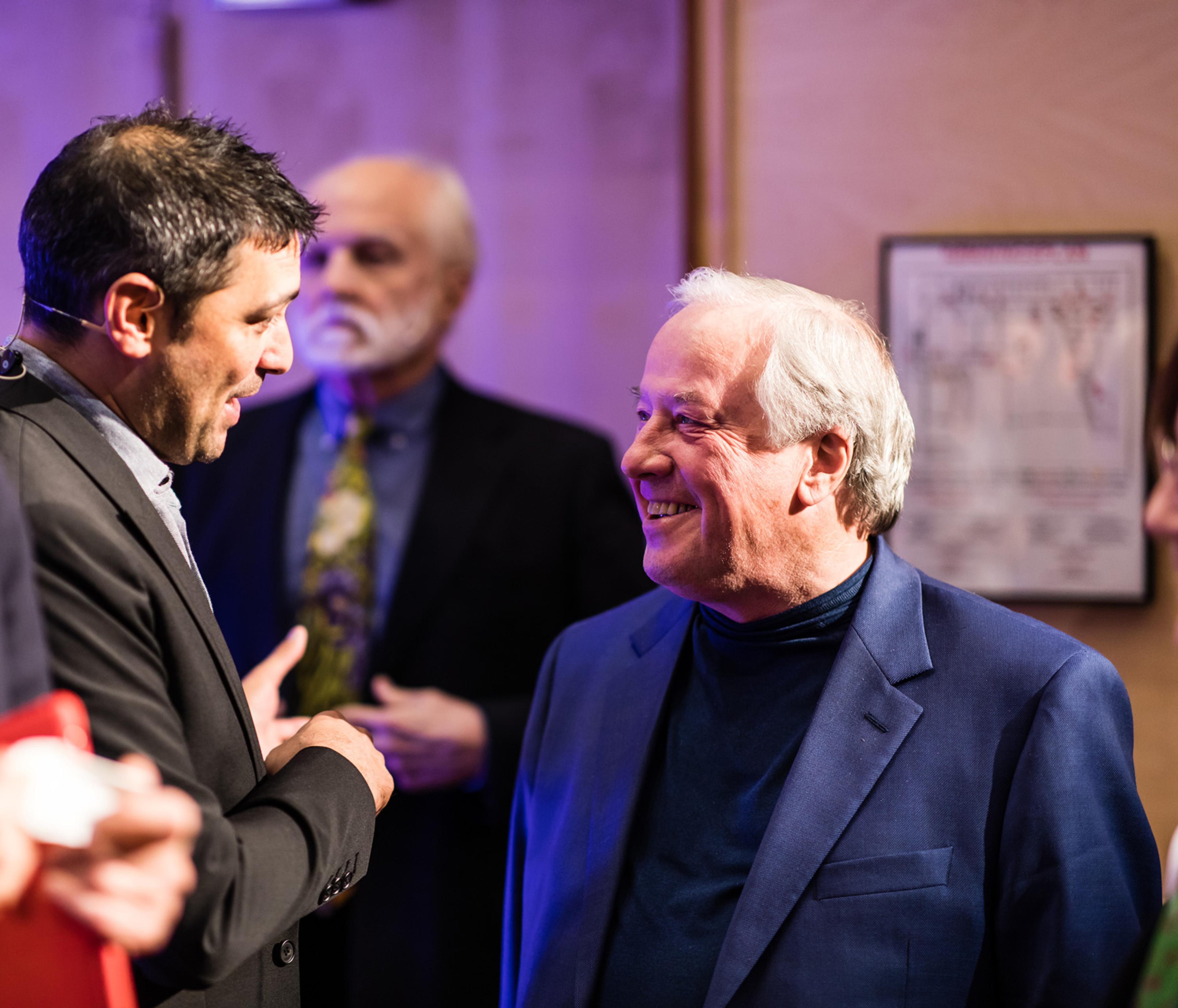
Robert Fettiplace in conversation with science journalist Adam Rutherford during the Kavli Prize week in Oslo (Photo credit: Thomas Eckhoff)
I was educated at Nottingham High School, a boy’s private school known as a direct grant school, that accepted a fraction of poor students funded by the local education authority. I was one of these, having been accepted based on my scores in the now defunct “eleven-plus” examination. It was an excellent school where I learned in detail all physical sciences (but not biology), mathematics and several foreign languages.
Favourite teacher
My favorite teacher was Mr. Pitts, a maths teacher, who encouraged and challenged me with many problems, including the famous four-color map problem (not finally proven until 1997); and mechanics problems, such as why moving bicycles do not fall over. My great grandfather and grandfather were both gamblers and off track bookmakers (illegal in England until 1960), which may account for my interest in mathematics!
Music
Besides mathematics, a preoccupation during this period was music. I had no formal musical education but, like many teenagers at that time, learnt to play the guitar influenced by Lonnie Donegan, Buddy Holly, the Shadows and ultimately the Beatles. I built my own imitation red Fender-Stratocaster electric guitar, and played in a rock group called ‘The Boys’ throughout school. My best friend was Alan Jones, a clarinetist, saxophonist and guitarist, whom I accompanied in several folk rock bands at college and later. I believe my passion for music partly fostered my later interest in the auditory system. The family appetite for music later continued with my son David, who was a cellist in the state youth orchestra; my son Michael, who, with his brother as bassist, was lead singer in a Madison rockabilly band; and my grandson Callum who currently plays bass in a jazz band.
Mathematical Physics
In 1965, I was accepted to Cambridge University to read Mathematical Physics, where I was initially taught by John Horton Conway, who invented the cellular automaton called ‘The Game of Life’. But mathematics seemed very dull, so in my second year, I switched to the Medical Sciences Tripos. This involved learning biology and taking a formal ‘A level’ biology exam, which was my first introduction to amazing topics such as cells, and genes, the brain and evolution, to fire my imagination. In my final undergraduate year, I took Physiology, taught at the famous Physiological Laboratory, with particular emphasis on neuroscience, graduating with an MA in medical sciences. The faculty in that department had included Nobel laureates Edgar Adrian, Alan Hodgkin and Andrew Huxley. Hodgkin, along with his student Huxley, elucidated the mechanism underlying the nerve action potential, and first proposed the existence of ion channels, protein pores that regulate flow of ions across the plasma membrane. He was a genius, the cleverest person I have ever met. He had a unique way of thinking about biological problems, which ones were subject to mathematical analysis and therefore potentially soluble, and which ones were not, and he had an enormous influence on my scientific thinking.
I continued as a research student at the Physiological Laboratory, investigating the structure and permeability of artificial lipid bilayers with Denis Haydon. In addition, I worked two seasons on the effects of channel-forming antibiotics such as the calcium-permeable nystatin on squid axons at the Plymouth Marine Biological Station. During my time at Plymouth, I met Denis Baylor and Andrew Crawford, both of whom later influenced my scientific career.

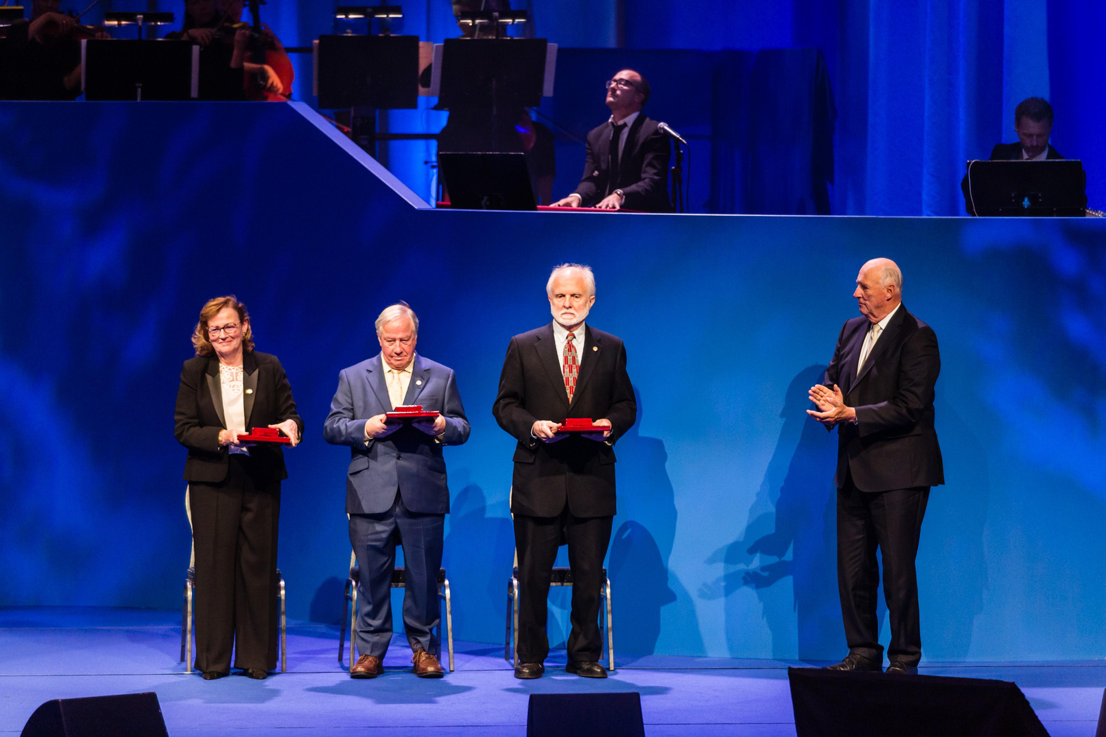
The 2018 Kavli Prize neuroscience laureates on stage in Oslo Concert Hall after having received the awards from His Royal Highness King Harald. From Left: Christine Petit, Robert Fettiplace, A. James Hudspeth and HRH King Harald (Photo credit: Thomas Eckhoff).
PhD in membrane biophysics
In 1974, after receiving a PhD in membrane biophysics, I joined Denis Baylor at Stanford University, where we worked on synaptic transmission in the turtle retina. This involved performing simultaneous recordings from photoreceptors (rods and cones) and ganglion cells. From these experiments, we determined that electrical signals of just a few millivolts in the photoreceptors could be transmitted through the retinal synaptic pathway to elicit action potentials in the ganglion cells. Furthermore, the pathway from rods had slower kinetic properties to the one from cones, so slow signals of time course comparable to the rod light responses were better conveyed via the rod pathway. Denis had previously produced four exceptional papers on photoreceptor transduction with Alan Hodgkin, and he handed on to me Alan’s inimitable experimental philosophy.
While at Stanford, I also met my future wife, Merriel Kruse, and returned with her to Cambridge in 1976. In searching for a new project at the Physiological Laboratory, I resolved that the field of photoreceptor research was too crowded and, furthermore, I decided, mistakenly as it turned out, that there was not much left to do in that area. I spent much time talking with Andrew Crawford, often in the Eagle pub in Cambridge, about what other research problems in sensory transduction to attack. At the time studying sensory receptors was considered a good inroad to exploring neural signaling, if only because it was possible to precisely control the stimulus, unlike the often used cerebellum or hippocampus, where the physiological inputs are largely unidentified. We turned to auditory transduction in hair cells, about which little was known.
There were rumors that the narrow frequency discrimination endowed by cochlear processing was vulnerable to lack of oxygen, suggesting that some active cellular mechanism contributed a ‘sharp second filter’. The obvious preparation to exploit was the turtle auditory papilla, because nerve cell preparations from this reptile could survive for a long time due to an ability for anaerobic metabolism. The robustness of the preparation had been recognized by Edgar Adrian, who, in 1938, recorded from the auditory nerves of both tortoise and alligator. Andrew Crawford and I developed an isolated turtle half head preparation with access to the auditory hair cells of inner ear, but where the middle and external ears were intact and could be stimulated with sound. We described the sound sensitivity and frequency selectivity of the hair cells, and discovered that each hair cell behaves as an electrical resonator, intrinsically tuned to a narrow frequency range set by its electrical properties. My work over the next decade, along with Jon Art and Paul Fuchs, demonstrated that the variation in resonant frequency along the turtle papilla (the tonotopic map) arose from systematic differences in the numbers and speed of calcium-activated potassium channels, and that the tuning could be modulated by stimulation of a cholinergic efferent synapse. In discovering electrical tuning, we believed we had stumbled across the ‘second filter’, but this proved incorrect. Electrical tuning was subsequently reported in auditory hair cells of amphibians, other reptiles and birds, but never in mammals. The most likely explanation for the difference is that evolution of small nocturnal mammals in a dinosaur dominated world required an expansion of the auditory range to frequencies too high for potassium channels to follow.
The upper frequency limit of hearing for most birds in 5 kHz, whereas for small primitive mammals, it can be 10 to 20 times higher. An alternative mechanism for a ‘second filter’ might be some kind of fast electro-mechanical process, and discoveries in this area emerged during the 1980s. One such process was the electrically-induced contraction of outer hair cells, first studied by Bill Brownell and Jonathan Ashmore, and extensively characterized by Peter Dallos and his colleagues, culminating in their cloning of the piezoelectric membrane motor ‘prestin’ in 2000.
A second process was active motion of the hair cell stereociliary bundle. Andrew Crawford and I first addressed this possibility by projecting images of stereociliary bundles on a pair of photodiodes, differentially recorded to follow rapid motion of the bundle with small nanometer-scale amplitude. Using this technique, we concluded that turtle stereociliary bundles possess an active force generating mechanism. We subsequently characterized active bundle motion in more detail in hair cells of turtles, chickens and rats. However, the mechanism was most clearly explained by the experiments of Jim Hudspeth and coworkers, who showed that active force generation was linked to activation and adaptation of the mechanically sensitive ion channels that normally transduce sound into electricity. Whether this process contributes in mammalian cochlear hair cells is still unclear.

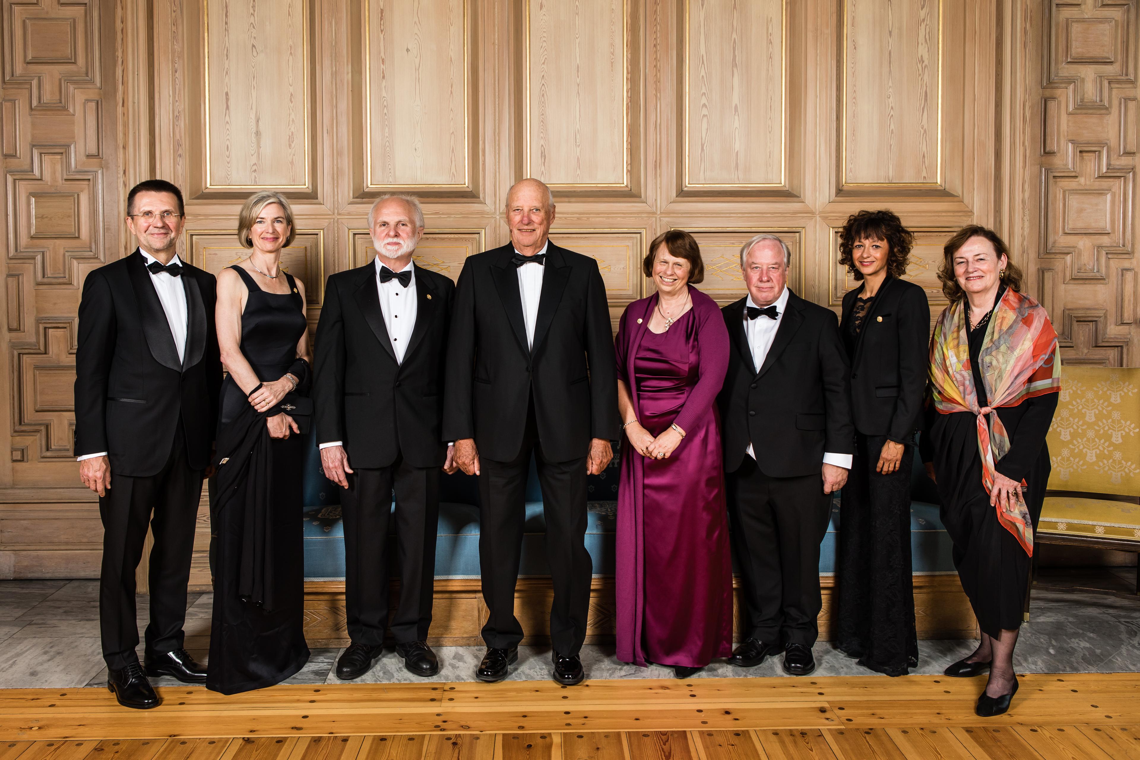
The 2018 laureates at the reception in the Munch room at Oslo City Hall, from left: Virginijus Šikšnys, Jennifer A. Doudna, A. James Hudspeth, His Royal Highness King Harald, Ewine van Dishoeck, Robert Fettiplace, Emmanuelle Charpentier and Christine Petit (Photo credit: Thomas Eckhoff).
Fellow of the Royal Society
In 1990, I was made a Fellow of the Royal Society of London, at an awe-inspiring ceremony, which involved signing a registry book containing signatures of all previous fellows, including Sir Isaac Newton. In the same year, I moved to the USA, partly to be closer to my wife’s family, and I became Steenbock Professor of Neural Sciences in the Department of Neurophysiology at the University of Wisconsin in Madison. The Department of Neurophysiology was distinguished for its achievements in auditory physiology, and contained researchers such as Jerzy Rose, John Brugge and Joe Hind, (who described auditory nerve phase-locking), Bill Rhode (first to measure the narrow tuning of the basilar membrane with the Mössbauer effect), Tom Yin (who described the brainstem sound localization pathway) and Donata Oertel (first produced tissue slices of the cochlear nucleus). My work during my time here has focused mainly on characterizing the properties, location and identity of the mechanically-sensitive channels, often referred to as mechanoelectrical transducer (MET) channels. Although the work was begun in turtle hair cells, I later switched to rat cochlear hair cells. Employing patch clamp techniques, we devised methods for isolating and stimulating single MET channels, which was very exciting since it revealed a tonotopic gradient in their properties. These ion channels are challenging to study because they exist in very small numbers and must be mechanically connected to other parts of the cell to function.
One of my most thrilling experiments was to use calcium-imaging to localize the MET channels. High speed video imaging, done in collaboration with Maryline Beurg and Tony Ricci at Stanford, provided unequivocal proof that the channels were at the tips of all but the tallest stereocilia, most likely in a complex demarcated as an opaque region in electron micrographs. This result constrained the method by which they were stimulated, but it gave no insight to their molecular identity. The study of mutations that cause deafness led to the elucidation by Christine Petit and others of many of the components of this protein complex within which the transducer channel is tethered. Until recently, however, it remained unclear which proteins form the channel. Discoveries of the effects of mutation by Andrew Griffith and colleagues at the NIDCD (National Institute on Deafness and other Communication Disorders) pointed to the transmembrane channel-like protein, TMC1, as a crucial element. My work has shown that the size of the single MET channel conductance, its variation along the tonotopic axis, its selectivity to calcium ions and its adaptive behavior are all modified in mutations of TMC1. This provides strong evidence that TMC1 forms the mechanoelectrical transducer channel.
One other line of research I have pursued since being in the USA is understanding the regulation of intracellular calcium, both as an important cytoplasmic messenger and as a potential source of hair cell damage. We were one of the first to apply real-time confocal microscopy to describe the spatial spread of calcium throughout the cytoplasm. This allowed me, in collaboration with Jon Art and Tom Tucker, to see calcium ‘hotspots’ around the synaptic release sites, and to characterize the mechanism of calcium extrusion via an ATP driven calcium pump. The importance of calcium turnover in hair cells is highlighted by the discovery that mutations of this pump, thus impairing calcium extrusion, underlie certain forms of deafness. Another indicator of the significance of calcium is the high levels of calcium binding proteins in the hair cell cytoplasm. In an extended collaboration with Carole Hackney, from Keele University in the UK, we documented the identities and concentrations of such calcium buffers. Carole was a superb electron microscopist who was one of the first to report tip links underlying force transmission to the MET channel. She used an ingenious method to quantify cytoplasmic proteins from their immunological labeling in electron micrographs. By measuring the density of the immune-gold particles in tissue sections and in gels containing known amounts of the protein under study, she could accurately determine their concentration in the cytoplasm. A surprising finding was that one particular protein, oncomodulin, related to the parvalbumin occurring in skeletal muscle, is present at millimolar levels in outer hair cells. It was recently shown that oncomodulin knockout mice display progressive hearing loss, consistent with a central role for calcium.
Different preparations and new techniques
Throughout my research, my aim has been to use different preparations and devise new techniques to study hair cells. The experiments have often been technically challenging, but have enabled us to be first to acquire different types of data. They were followed up by later quantitative analysis and coupled with modeling to complete the story. However, the results of electrophysiological experiments are usually evident immediately, which can be exciting to experience. It makes one feel like an explorer who turns a corner in the forest to encounter a new and unexpected artifact. Furthermore, many such experiments have involved only one or two other people working at the equipment rig at the same time. While in the USA, I have been particularly fortunate to work first with Tony Ricci and more recently with Maryline Beurg, who are both good friends.
Fellow of the American Academy
My research was rewarded with election as a Fellow of the American Academy of Arts and Sciences, the oldest learned society in USA, founded in 1780, not much later than the Royal Society. I was accompanied to the election ceremony in 2012 by my son, Michael, who has become a physician and is practicing at Massachusetts General Hospital, and sharing the occasion with him made me feel very proud.
For over 20 years at the University of Wisconsin, I have enjoyed teaching a neuroscience course attended by both undergraduates and graduate students. The students are so enthusiastic about neuroscience. Interactions with them
and with my colleagues persuade me that I can still make a valuable contribution, and that keeps me sufficiently young to continue with the laboratory research.