


Changing Our View of the Nanoscale World
A Conversation with Gerd Binnig and Christoph Gerber
The 2016 Kavli Prize laureates discuss how the ability to see and manipulate single molecules and atoms has changed our view of the nanoscale world.
View of cantilever in Atomic Force Microscope [magnification 1000× ; resolution ~ 0.1 μm per pixel] (Credit: SecretDIsc, Wikimedia Commons)
By Alan S. Brown
Thirty years ago, Gerd Binnig, Christoph Gerber and Calvin Quate began developing a device that would enable us to see features smaller than one nanometer–less than 1/50,000 the diameter of a human hair and far smaller than any traditional microscope could manage. Since then, their atomic force microscope, or AFM, has become one of the most important tools for understanding the nanoscale world. Researchers have used it for such wildly different tasks as unfolding proteins, watching chemical reactions as they occur, and arranging atoms to probe their quantum properties.
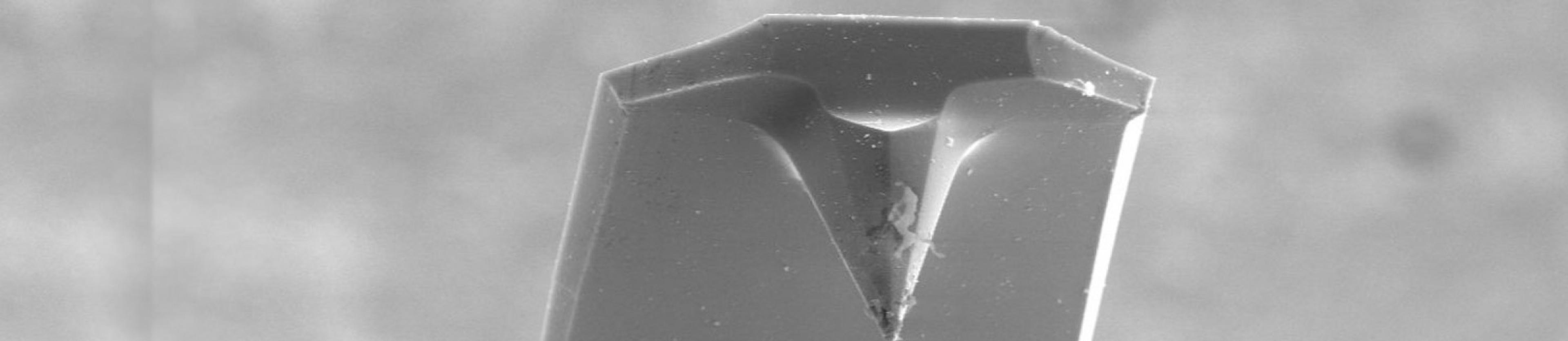
This breakthrough device hinged on the ability to measure the forces exerted by individual atoms. The AFM did this with a design that very much resembled a record player. Both consist of a long arm, or cantilever, with a fine tip at the end. On a record player, the tip vibrates as it traces the grooves in a record, and electronics translate those vibrations into sound. On the AFM, the arm and tip are so fine, that the tip moves when attracted or repelled by individual atoms.
For the invention and realization of atomic force microscopy, Binnig, Gerber and Quate received the 2016 Kavli Prize in Nanoscience. During a teleconference roundtable, the laureates discussed the joys of hard problems in science, and how AFM continues to change the way we see the world.
Our roundtable panelists were:
- GERD BINNIG –is a physicist and Nobel Laureate for his invention (with Heinrich Rohrer and Christoph Gerber) of the scanning tunneling microscope while at IBM Zurich. He began development of the atomic force microscope in 1986 to overcome the limitations of his previous invention.
- CHRISTOPH GERBER –is a physicist and director for scientific communication at the Swiss Nanoscience Institute at the University of Basel. While at IBM, Gerber worked closely with Binnig on bringing both the scanning tunneling microscope and atomic force microscope to fruition.
Calvin Quate was unable to participate in the roundtable. The transcript has been amended and edited by the laureates
You filed your first patent for the atomic force microscope (AFM) nearly 30 years ago. How has it changed the way we look at the world since then?
GERD BINNIG: It was like the first time people looked through an optical microscope and saw bacteria. That completely changed how we look at the world. Suddenly, we understood what was really going on in nature, and we used that knowledge to learn how diseases spread. The AFM is the next step. It lets us look at the molecules that make life possible in those bacteria – and everywhere else – and see things we could not see before. It teaches us how to make changes to surfaces or molecules that we attempted blindly in the past. And it has been used in so many different scientific studies, from looking at polymers and chemical reactions to modifying surfaces at the atomic level.
CHRISTOPH GERBER: As Gerd explained, seeing is believing, and now we can do that onthe atomic scale. AFM has turned into the most powerful and most versatile toolkit that we have for doing nanoscience. And it keeps evolving. In just the past few years, researchers have learned to pick up a molecule on the tip of an AFM, which we can think of as the needle on a record player, and reveal chemical bonds while imaging molecules on surfaces. Nobody thought that ever would be possible.
Has this changed how researchers think about the ways nanoscale interactions affect the things they study?
BINNIG: Very much so. Before AFM, people who wanted to model very small structures –molecules, cell walls, semiconductors – had to make indirect measurements of them. But those structures can be complex and disordered, and indirect measurements do not always capture that, so the models they came up with were often wrong. But now, we can look at those structures and adapt our models to match what we observe. We as scientists always have to connect our theories to reality. Atomic force microscopy lets us do this.
When you started thinking about the AFM, biology was one of the fields you had inmind. Yet even you must have been surprised at how it has revolutionized biology.
GERBER: Yes. AFM’s capabilities keep evolving, and researchers are always finding new ways to use it. For example, in recent years, researchers have made tremendous progress in taking AFM measurements in real time. It’s like watching a movie. They can now see biological interactions, such as how molecules degrade or how antimicrobials attack bacterial membranes as they occur – something nobody could have foreseen 20 years ago. It took 15 years to get there, but we can now see biology in action and compare that to our theories.
BINNIG: Exactly. In biology, the biggest and most important question is always whether a molecule will bind to another molecule, change it, and by changing it cause something important to happen. This is all about forces, and researchers can use AFM to bring two molecules or even two cells close together, or pull them apart, and measure those forces directly. We can learn how big those forces are and under what conditions they occur. We're actually looking into the heart of biology when we do that.
GERBER: And atomic force microscopy can tell us about many different types of forces that determine the outcome of chemical reactions at the nanoscale. These range from chemical, mechanical and electrostatic through, most recently, to the very weak interactions between molecules.
BINNIG: A great example of this is how Hermann Gaub, a professor of biophysics at Ludwig Maximilians University of Munich, used AFM to unfold proteins. He actually attached one end of a protein to a surface and the other end to an AFM tip. When he pulled the tip up, the protein straightened out and he could create a fingerprint of the unfolding forces that he could compare with his model.
And does a more accurate model of how proteins are structured help design better medicines?
BINNIG: Exactly. In the new field of immunotherapy, for example, we want to use molecules to bind to the surface of an immune cell and activate that cell so it attacks cancer. To do this, we need to know which molecules bind to the right molecule on the immune cell. So we're back to the essential question, bind or not bind, and accurate models can help us answer that.
GERBER: And that's what I mean when I say that AFM has developed into this very versatile toolkit.
Has that surprised you? You developed the microscope to measure surfaces at the atomic scale. Today we use it to push around atoms, quantify molecular forces and image high-speed chemical interactions. This has made it possible to measure the attraction between a protein and a cell wall, or construct devices for quantum computing. Did you see any of this coming?
GERBER: Our sole idea in developing AFM was to image nonconductive surfaces, such as ceramics or biological materials, at atomic resolution by scratching a very fine needle on a long arm, or cantilever, over that surface. It was hard to foresee how people would pick up on this very simple idea to invent all these wonderful ways to answer difficult questions. And it happened so quickly. We came up with a very precise way to measure forces with that cantilever, and within just two or three years, someone figured out how to use a laser to get even better resolution. It took only five years until the first commercial instrument, and from there, there was no holding it back.
What about you, Dr. Binnig? Were you surprised that scientists could manipulate something as small as an atom?
BINNIG: Not really. In principle, once you can observe something with a tool, you're usually in a position to change or modify it. A laser, for example, can measure a surface, but turn up the power and you destroy it. The same is true of AFM. In principle, this is something we thought about from the very beginning.
What about applications you could not have foreseen?
BINNIG: I could not have foreseen that we can image molecules with such a high resolution. It's unbelievable. We can see the bonds between molecules. We can watch them change during a chemical reaction, and sometimes there are surprises. Some researchers have observed an intermediate state in a chemical reaction that should not have lasted long enough to see. So they have had to rethink their theories to take into account why this intermediate state lasted so long. That's what happens when we can observe such high-resolution details.
GERBER: Another example is high-speed AFM, which biologists use to see the cellular machinery in action. No other technique can do that. It works by tapping a very, very thin cantilever up and down, taking one quick measurement after another.
BINNIG: It is amazing how many people use the AFM in so many different fields. We first thought, well, maybe biology or semiconductor research. But it was picked up everywhere, from studying friction to cosmetics.
GERBER: I recently looked it up, and AFM was mentioned in 353,000 peer-reviewed papers. Our original article was published in Physical Review Letters, the top journal in the field in which all the important theoretical work is published. Ours is the only experimental paper on its list of most-cited papers.
Amazing. And yet AFM was actually a follow-up to another technology you worked on, the scanning tunneling microscope, or STM. It was probably the first instrument to achieve nanoscale resolution without using electrons or other high-energy beams that can damage what you are observing, right?
BINNIG: Yes.
And where did that idea come from?
BINNIG: We were trying to solve a problem. IBM was working on a new type of semiconductor chip, and the insulator, which keeps the electric current from escaping the semiconductor, was leaking. But no one knew why. So Heinrich Rohrer, who was working at IBM Zurich, hired me. I looked to all the available instruments, and none of them could study materials on such a fine scale to find out.
So the two of us thought, well, okay, we'll invent something. We thought we could take advantage of something called quantum tunneling. Quantum tunneling is when an electron tunnels through a conducting material and come out the other side. We developed STM to map the surface of the material by measuring where electrons emerged on the other side. Only later did we realize that we could move our probe from one spot to cover the entire surface.
Dr. Gerber, you quickly became part of the STM team. What convinced you to join?
GERBER: I felt this was such a crazy idea, and I'm always very fond of this sort of thing. I thought this was fantastic.
BINNIG: I can confirm this. Christoph always likes crazy things. That runs through his life.
GERBER: Actually, the development of STM was kind of an undercover project at the beginning, because Gerd and Heinrich were involved in other projects. I worked for a year or so on my own. When we started overcoming problems and we could see features on the surface of a material that were one-tenth of a nanometer, then it really took off.
BINNIG: Once you have the idea, it's mainly technical problems that remain. We needed a team, and Christoph joined, fortunately. His very optimistic view of the world and his way of dealing with problems made him a very important member of this group.
GERBER: Thank you, Gerd. I’ll take the flowers. I mean, it was fantastic. Those were high times when we succeeded, and that was really an unbelievable time in my life.
"Seeing is believing, and now we can do that on the atomic scale."—Christoph Gerber
Dr. Binnig, you and Heinrich Rohrer won a Nobel Prize for the scanning tunneling microscope in 1986. How did that change your research?
BINNIG: For some time, I actually stopped working. I helped people apply the technology, gave presentations and traveled around the world many times. But after a while, I wanted to go back to the lab. I learned to be really cruel and say, "No." So it was painful because people didn’t like that, and would come back to me again and again. But then the Nobel Prize got helpful, because I could get more support for a project, or ask other scientists for help from any place in the world.
How about you, Dr. Gerber? You were also part of the STM team.
GERBER: Heinrich and Gerd deserved it. Gerd, for me, is a genius, and I've never worked with anybody in my life that has such high standards.
BINNIG: Now I'm taking the flowers.
GERBER: I mean it, you know. He has a crazy mind, you see, and that's why I like it so much. Gerd can anticipate things, even if they are not in his field, and he is so quick in understanding what's happening. In America, you would say, “He’s awesome!” We worked together for 10 years, and I have to admit that it was the most fruitful and fantastic time in my life.
BINNIG: Those 10 years were fantastic, and they included our work on the AFM.
So let's get back to the atomic force microscope. Its predecessor only worked with conductive materials, such as metals and semiconductors. That was a problem, so how did you come up with a way to see living organisms, biomolecules, plastics, ceramics and a lot of other materials?
BINNIG: I thought, in principle, why do we need to have an electrical current and measure electrons? Why can't we just measure the forces between an atom sitting on a surface and an atom sitting on the tip of a probe? Physically, it would look a lot like what we were already doing with scanning tunneling microscopy. The question was whether it was possible to measure something as small as the forces between two atoms.
This was in 1986 and, both of you had moved to California. Dr. Gerber, you were at IBM's Almaden Lab in San Jose. Dr. Binnig, you were working at nearby Stanford University with Calvin Quate, the third recipient of this year’s Kavli Prize in Nanoscience. How did that come about?
BINNIG: I was actually working quite intensively with Calvin Quate's group. He invited me to California and gave me a visiting professorship for one-and-a-half years.
GERBER: Cal Quate had an interesting background. He was also working on microscopes, and had developed a way to use sound to achieve much better resolution than any optical microscope. But when he heard about the scanning tunneling microscope, he flew to Switzerland to visit us, and then redirected his group to begin looking at scanning tunneling microscopy. So we were very well acquainted with him and his work.
BINNIG: I have to say that he put together a wonderful group. He was a perfect scientific manager. The atmosphere he created in his group was something I've never seen before at a university. If one of the team members discovered something or got some nice results, all the others gathered around and wanted to know everything about it. They were all really happy for this one person. There was no jealousy. Zero, zero jealousy. Just a real team. I've never seen something like that again.
GERBER: It was just a fantastic group. And they worked in what is presumably the more American way of doing things, taking on high-risk stuff. They liked that. The European way of looking at things is usually more cautious.
So Professor Quate was also doing advanced work in atomic-scale microscopy. Did you initially discuss your ideas about the AFM with Professor Quate?
BINNIG: Yes, and with Christoph, too. We concluded that maybe it was actually doable, so Christoph and the rest of us started to build a system.
So you had this great idea. How did you turn your concept into reality?
BINNIG: We needed to build a cantilever, so Christoph and I went to a music store and bought a diamond-tipped record player needle. We took it back to the lab and smashed the tip with a hammer to get these tiny specks of diamond. We glued one of them on top of a very thin gold foil to create a little cantilever. That was the first atomic force microscope. Soon after that, Cal Quate and his team used advanced fabrication techniques to make a very sharp tip that came to a very, very small point.
GERBER: Yes, using Quate’s tip gave us the first hint that we could get atomic resolution, though it was not yet true atomic resolution. That came later.
Not many universities had the ability to machine such fine parts in the 1980s, did they?
BINNIG: Cal had good foresight and understood that things had to get smaller, and he was actually building scanning tunneling microscopes on semiconductor chips when we began working with him.
GERBER: Exactly. Cal had built up his capabilities to make very small devices. This enabled him to build AFM arms, which must be thin enough to bend and have a sharp tip to measure atomic forces.
There were so many technical challenges. Did you ever get frustrated?
GERBER: When it didn't work, we went out and played a round of golf.
BINNIG: That's true. Actually, we never got too frustrated with each other or the rate of progress because so many exciting things were happening. There were moments when everything went wrong, but that's part of the game. It's like soccer. When somebody scores against you, it's frustrating for the moment. But then you try hard to turn the game around. It's also like that in science.
GERBER: Gerd knows this because we had a soccer team at IBM, and he was the most gifted forward on this planet. He could use both his legs equally well, and this was mindboggling because his opponents never knew which way he would pass. He was fantastic. We did all kinds of things beside trying to do very good science, you know. It was a fantastic time.
Did you find those breaks helped when you were struggling with a problem?
BINNIG: When I'm running or playing football, I'm focused on something. But sometimes, when I'm just doing nothing, lying on the couch, for instance, that's a creative situation because I'm not focused on anything. The ideas flow by themselves because I don't have control. That is a good time to come up with something new, at least for me.
How about you, Dr. Gerber?
GERBER: Sometimes that's true, but other times I get ideas when I really work at it. Working on both instruments, the STM and the AFM, was really a day and night job. We spent so many nights in the lab it was unbelievable, but we had to do it to find a way forward. You know, there are some people, in my opinion, who are afraid of their own courage. They have this great idea, but they are maybe afraid to do the step-by-step work to prove their principles. Yet they still try to turn their idea into a Ferrari, and are surprised that it doesn't work. By proving those principles systematically, I was better prepared to struggle with the inevitable technical problems.
"We as scientists always have to connect our theories to reality. Atomic force microscopy lets us do this." — Gerd Binnig
Clearly, your work on the atomic force microscope changed how other scientists explore and understand everything that happens at the nanoscale. How about yourselves? Are you still working with AFM or are you doing different things now?
BINNIG: I like to do different things from time to time. So 16 years ago, I founded a company that develops software that makes computers a little bit more intelligent. We call it e-cognition, and it is similar to what IBM calls cognitive computing. Our software automatically analyzes medical images. Specifically, we have the computer look at tissue samples from cancer patients and try to predict the best kind of treatment. So in this respect, I'm doing something similar to Christoph, who also moved into medicine. Though I am planning to retire soon, and so maybe I will move back to AFM.
GERBER: Hopefully.
BINNIG: I'm thinking about it, yes, I'm really thinking about it.
GERBER: It's about time, Gerd, it's about time.
BINNIG: Yeah, we'll be doing something. I missed you.
And how about you, Dr. Gerber?
GERBER: My group at the Swiss Nanoscience Institute is using AFM to study mutations of cancer patients in order to improve diagnosis and treatment. Our system uses arrays of AFM to scan RNA, the chain-like molecule that translates DNA into proteins, and look for mutations, or even fractional mutations where only an atom or two are in the wrong positions. Doctors can use this information to plan treatments for their specific type of cancer. It took us 16 years to get this technology into clinical trials, but we can make our diagnosis in less than one day compared with existing methods that take more than one week.
BINNIG: So, in principle, Christoph and myself, we ended up in the same spot. We started with atomic-scale imaging and now we are both trying to diagnose cancer patients. It's quite a coincidence.
So, final question. It's been 30 years since you started working on AFM. In all that time, what is the most surprising thing you've learned?
GERBER: It goes back to what I said before, the AFM keeps evolving. The atomic resolution, the ability to see atoms and the bonds between them, the ability to see chemical reactions happen in real time – this is all just simply fantastic. It just keeps getting better and better.
BINNIG: For me, there are two aspects. One is that AFM is used so widely in so many different fields. I never imagined that would happen. And two, as Christoph says, it keeps getting better and better, and shows us more and more. That, I think, is a surprise to me.