


Exciting Challenges
As told by Brenda Milner
Since 1950, the bulk of my research has been carried out at the Montreal Neurological Institute, where the availability of patients undergoing unilateral removals from temporal or frontal lobes to control epileptic seizures provided a unique opportunity for studying functional representation in the human brain. The research has evolved around three main themes; the complementary specializations of the human cerebral hemispheres; the role of the medial temporal-lobe structures, and particularly the hippocampus, in the establishment of new long-term memories; and the cognitive changes associated with frontal-lobe lesions. In addition, this long series of neurosurgical patients tested has allowed me to study speech lateralization preoperatively in lefthanders and right-handers using the Wada sodium-amobarbital procedure and to explore the effects of early unilateral brain lesions on cerebral organization of language at maturity. I have also adapted the Wada technique to screen patients for possible risk to memory in a planned temporal lobectomy.

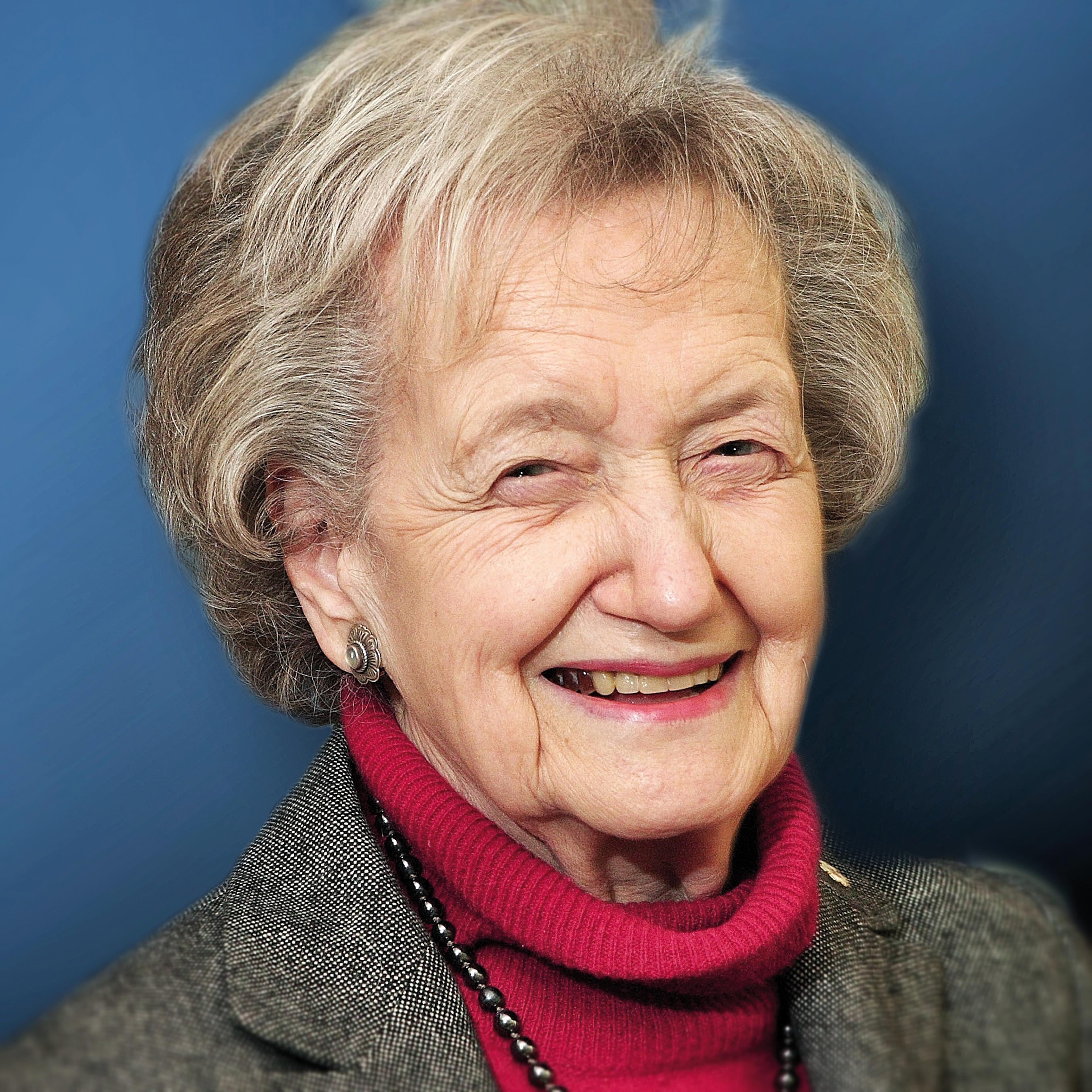
Brenda Milner (Photo credit: © Owen Egan/McGill, University).
Temporal-lobe studies: effects of unilateral lesions
Influenced by Klüver and Bucy’s findings in the monkey, I looked first for perceptual deficits on complex visual tasks, finding mild impairments after right temporal lobectomy, but not after left. Much stronger results emerged when I switched to memory tasks, where I found clear and complementary material-specific deficits related to the side of the lesion: verbal from the left, and non-verbal from the right, with impaired memory for faces, abstract drawings and simple melodies being observed after right temporal lobectomy but not after left.
The medial temporal-lobe amnesic syndrome
The deficits described above were mild and material-specific; in contrast, in two patients, PB and FC, a left temporal lobectomy that included the hippocampus resulted in a generalized anterograde amnesia for the events of daily life, together with a retrograde amnesia for the preceding few months, but unaccompanied by other cognitive deficits. To account for this unexpected memory loss after a unilateral removal, we speculated that in each case there had been a pre-existing but preoperatively unsuspected atrophic lesion in the hippocampal region of the opposite hemisphere, so that the operation effectively deprived the patients of hippocampal function bilaterally. We emphasized the hippocampal region because PB had had his temporal lobectomy in two stages, five years apart, and it was only after the medial removal that the memory loss occurred. Our hypothesis was confirmed 12 years later when autopsy findings on PB revealed longstanding right hippocampal atrophy.
Our report of these two cases attracted the attention of Dr. William Scoville, a neurosurgeon from Hartford CT, who told us that he had observed a similar memory disturbance in a 27-yearold patient on whom he had performed a bilateral medial temporal-lobe resection, also in an attempt to control epileptic seizures. As a result I was invited to go to Hartford to study the now famous patient HM.
The challenge was twofold: (1) to analyse the nature of the memory impairment and (2) to determine whether any new learning was possible. As with our Montreal patients, HM’s immediate memory, a limited-capacity system, was intact, and simple verbal items could be retained across many minutes by constant rehearsal. But if rehearsal was prevented, or if the material could not be easily verbalized (as with lights flashing at a particular frequency, or sounds clicking), then HM was unable to maintain the mental representation required to bridge the gap. It was not until 1976 that Mishkin corroborated these findings for the monkey, using a delayed non-matching to sample task, thus providing a convincing animal model for HM’s amnesia.
HM’s failure on single-trial memory tasks accorded with his forgetting of the events of daily life, but it did not rule out the possibility that he might be capable of some kind of learning, given extended practice. Here I had my major breakthrough in showing completely normal learning on a sensorimotor task (mirror drawing) over a 3-day period, with HM amazed at his own success, since he was completely unaware that he had done the task before. This study was important in providing early evidence for the existence of more than one memory system in the brain. We now know that motor-skill learning depends critically on the basal ganglia.

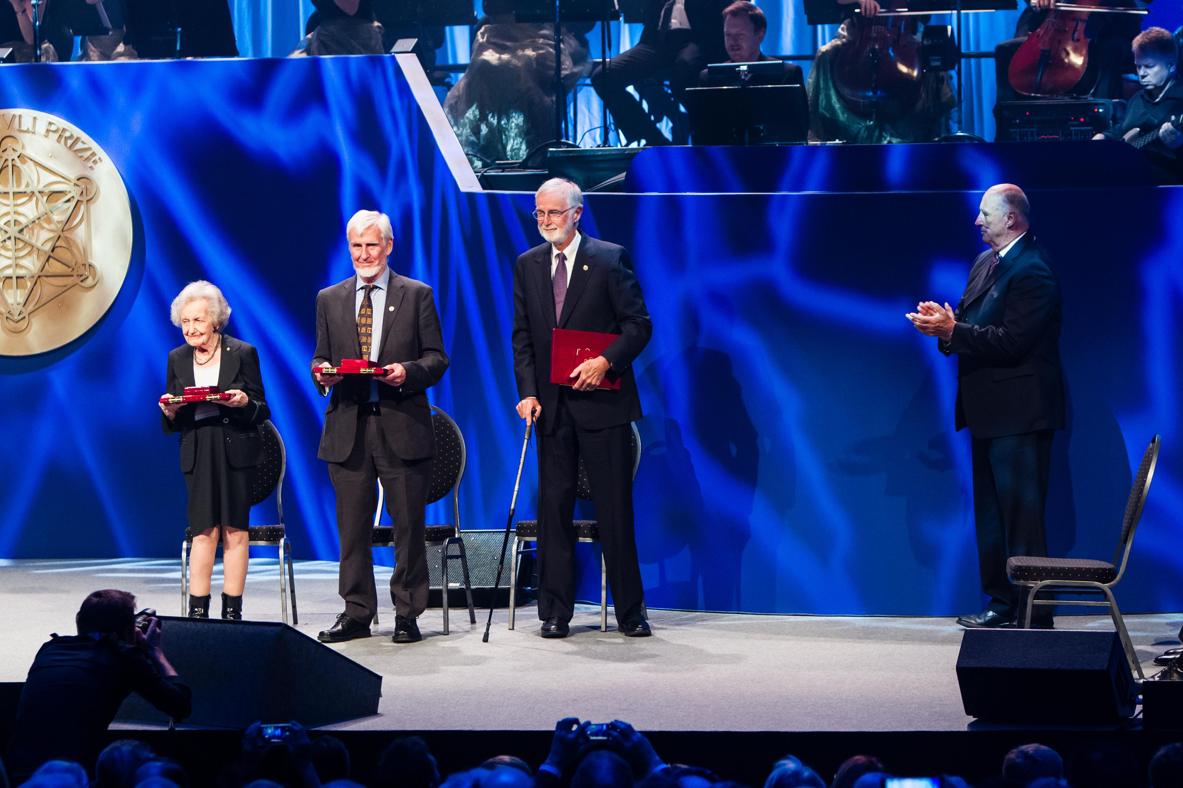
All three 2014 Kavli Prize laureates in neuroscience on stage at the ceremony is Oslo together with His Royal Highness King Harald. From left: Brenda Milner, John O'Keefe and Marcus E. Raichle (Photo credit: Scanpix).
Frontal-lobe studies
Strange as this may seem today, the importance of the frontal lobes for cognitive function was in serious question in the 1950’s, largely owing to the good performance of many patients with large frontal-lobe lesions on standard intelligence tests. This led Hebb and others to suggest that the frontal lobes may be important for intellectual development but that this region played a less critical role in the adult brain. Indeed, my own frontal-lobe groups performed normally on most of my cognitive tasks. But, guided again by work in the monkey, I used the Wisconsin Card Sorting Test to elicit strikingly perseverative behaviour in patients with dorsolateral frontal-lobe lesions, particularly in the left hemisphere. Despite initial scepticism, this task is now used widely in clinical neuropsychology as a possible indicator of frontal-lobe dysfunction.
Fluency tasks: a measure of divergent thinking
Traditional intelligence tests may be said to measure different aspects of convergent thinking, in the sense that there is usually just one answer to the question or problem set. Tests of divergent thinking, in contrast, emphasize the number and variety of answers that can be generated to a single question. Such tasks are held to be better predictors of creative achievement that are standard intelligence tests and we found evidence that performance on such tasks may be particularly vulnerable to the effects of frontallobe injury. Our first evidence came from the use of the Thurstone Word Fluency task, and impairment being found after left frontal lesions in patients who were performing normally on other verbal tasks. This task requires the subject to list as many words as possible beginning with a particular letter within a short time limit. A corresponding deficit on a nonverbal fluency task when subjects were asked to draw as many different unnameable designs as they could invent in a given time. Here the right frontal-lobe group was more severely impaired than the left, another instance of complementary functional specialization.

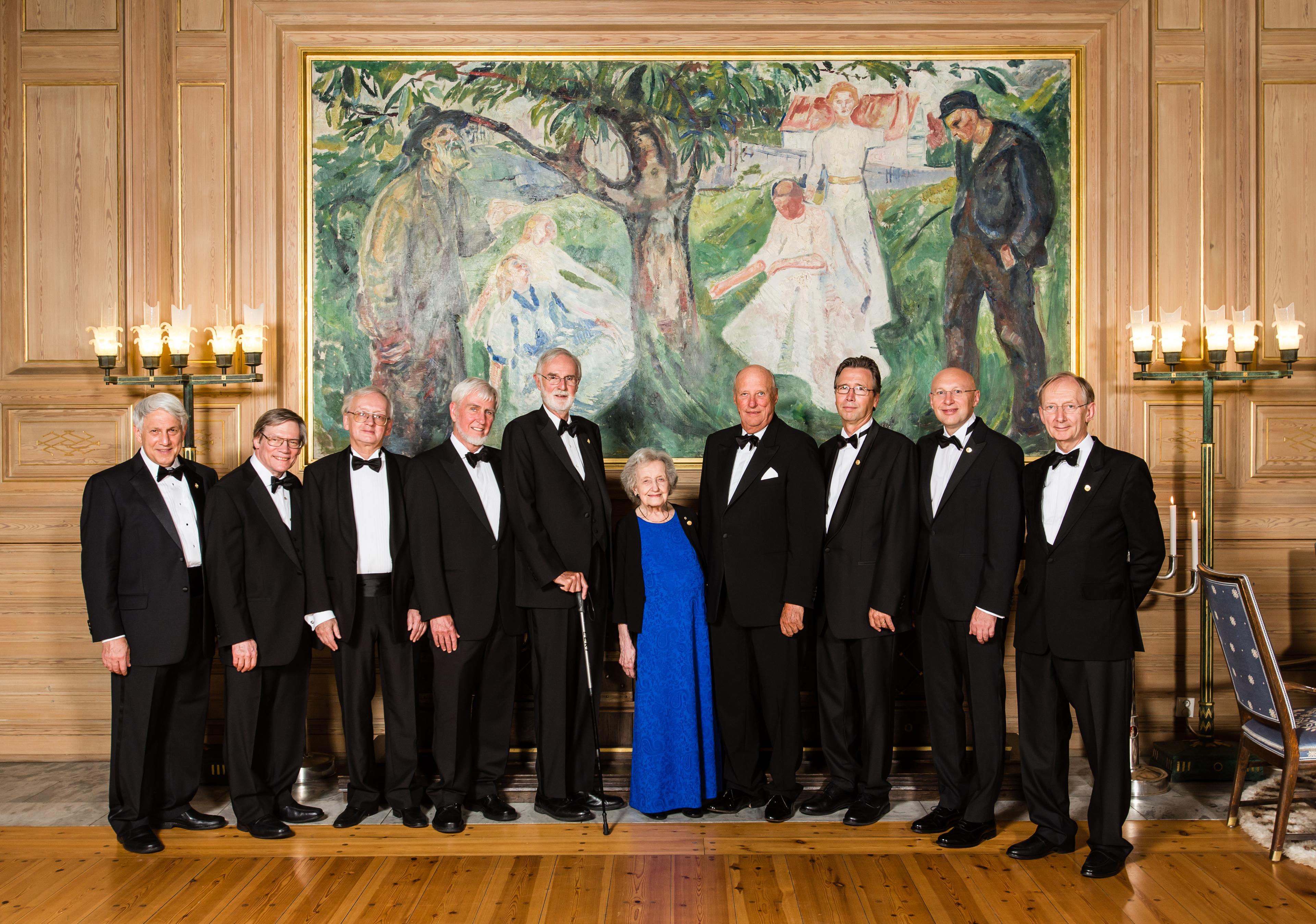
All the 2014 Kavli Prize laureates together with His Royal Highness King Harald in the Munch room at Oslo City Hall at the reception prior to the Kavli Prize banquet. From left: Andrei D. Linde, Alan H. Guth, Alexei A. Starobinsky, John O'Keefe, Marcus E. Raichle, Brenda Milner, His Royal Highness King Harald, Thomas W. Ebbesen, Stephan W. Hell and Sir John B. Pendry. (Photo credit: Thomas Eckhoff).
Frontal lobes and the temporal organization of memory
A scrutiny of which memory tasks proved difficult for frontal-lobe patients, and which did not, led me to propose that frontal-lobe lesions might interfere with the ability to structure and segregate events in memory, and hence that, in a situation lacking strong contextual cues, patients with such lesions would be less able than normal subjects to give salience to a stimulus that had been presented 60 seconds ago over one that had appeared earlier in the same series of trials. In this case, a task that specifically requires temporal-order judgments for items presented in a series should prove difficult for patients with frontal-lobe lesions. We have since confirmed this hypothesis using three recency-discrimination tasks, with contrasting effects depending on the side of the lesion and the nature of the stimulus material. How far the impaired performance of patients with frontal-lobe lesions represents a failure to develop appropriate encoding and retrieval strategies rather than the breakdown of an automatic time-tagging process remains to be determined.

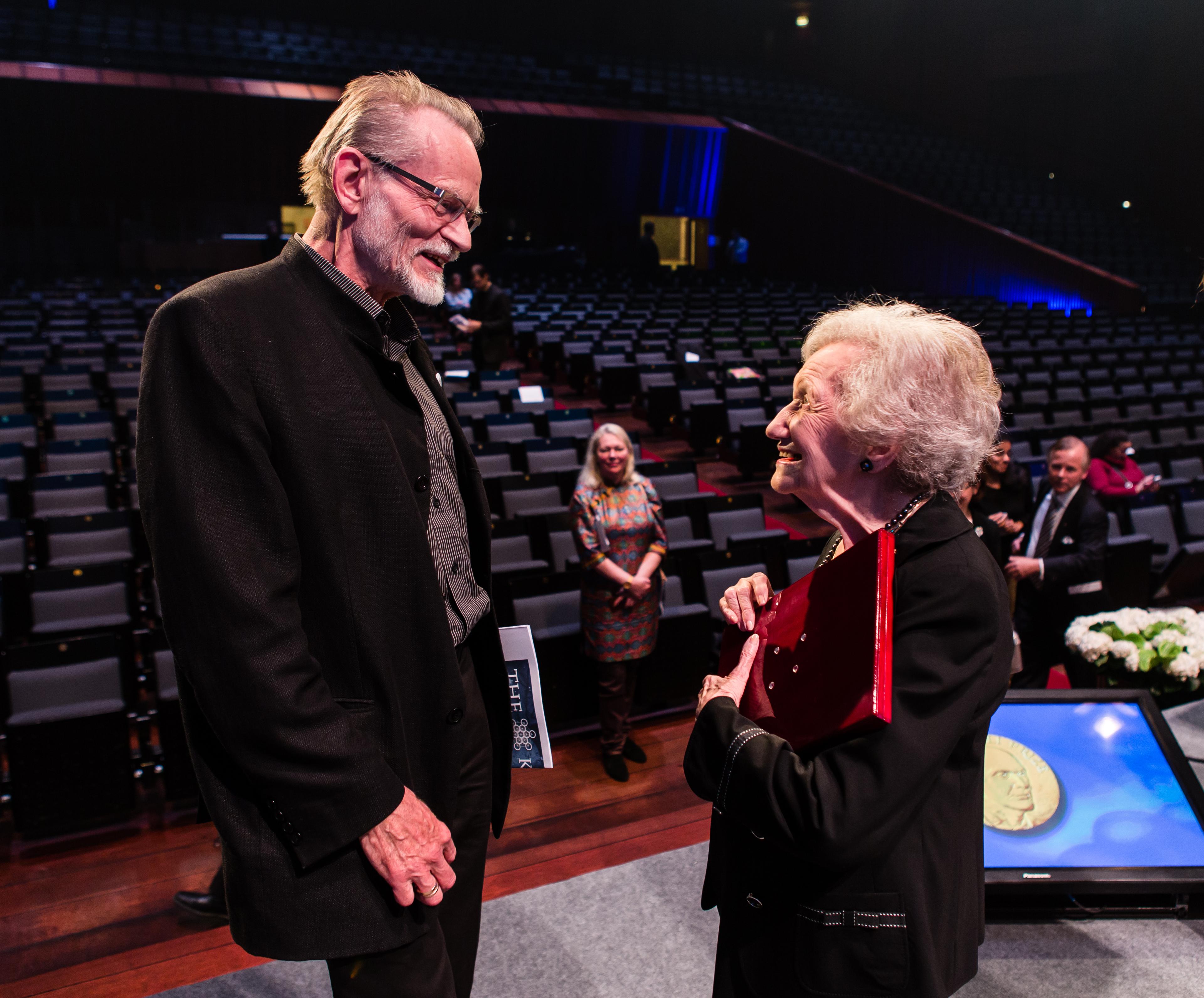
Brenda Milner in discussion with the chair of the Kavli Prize neuroscience committee 2008-2012 Jon Storm-Mathisen (Photo credit: Scanpix)
Comment
What these three stages of my work have in common is an empirical approach. The research has been driven by observed curiosities of behaviour that I felt the need to explore with appropriately designed tasks. Many of the findings ran counter to prevailing views of the day but they have stood the test of time, as well as stimulating a whole new experimental interest in the field of brain and memory. Meantime, new technology leads the way, and it is now possible to use functional neuroimaging techniques (PET and fMRI) to observe the normal brain in action during the performance of cognitive tasks. I have recently collaborated with Denise Klein to investigate language representation in normal bilingual subjects, and, with Joelle Crane, I am using structural magnetic resonance imaging to explore the contribution of different medial temporal-lobe structures to object-location memory. Thus, we are learning more and more about hemispheric specialization. The exciting challenge is to understand better how the left and right hemispheres work together, in health and in disease.