


Discovering New Features of the Brain's Organization and Function
As told by Ann Martin Graybiel
I was born in Boston, Massachusetts, but was raised in Pensacola, Florida, a then-small town in the north of Florida. My father had been sent there from Boston during wartime. He was a cardiologist and research scientist; my mother, from Northern Ireland, was herself from a medical family. Both were enormously influential in my development. I was privileged to learn about the excitement of science from my father, and from both parents the importance of medicine and of giving through one’s life-work to society. Schools at that time were still under-developed in that region (for example, being a girl, I was not allowed to take a science course — girls had to take sewing class). My parents sent my brother, Ashton, now a physician, and myself to boarding schools. At the National Cathedral School in Washington DC, I reveled in the music at the Cathedral, and in the arts and history to which we were exposed. I decided to become an historian.

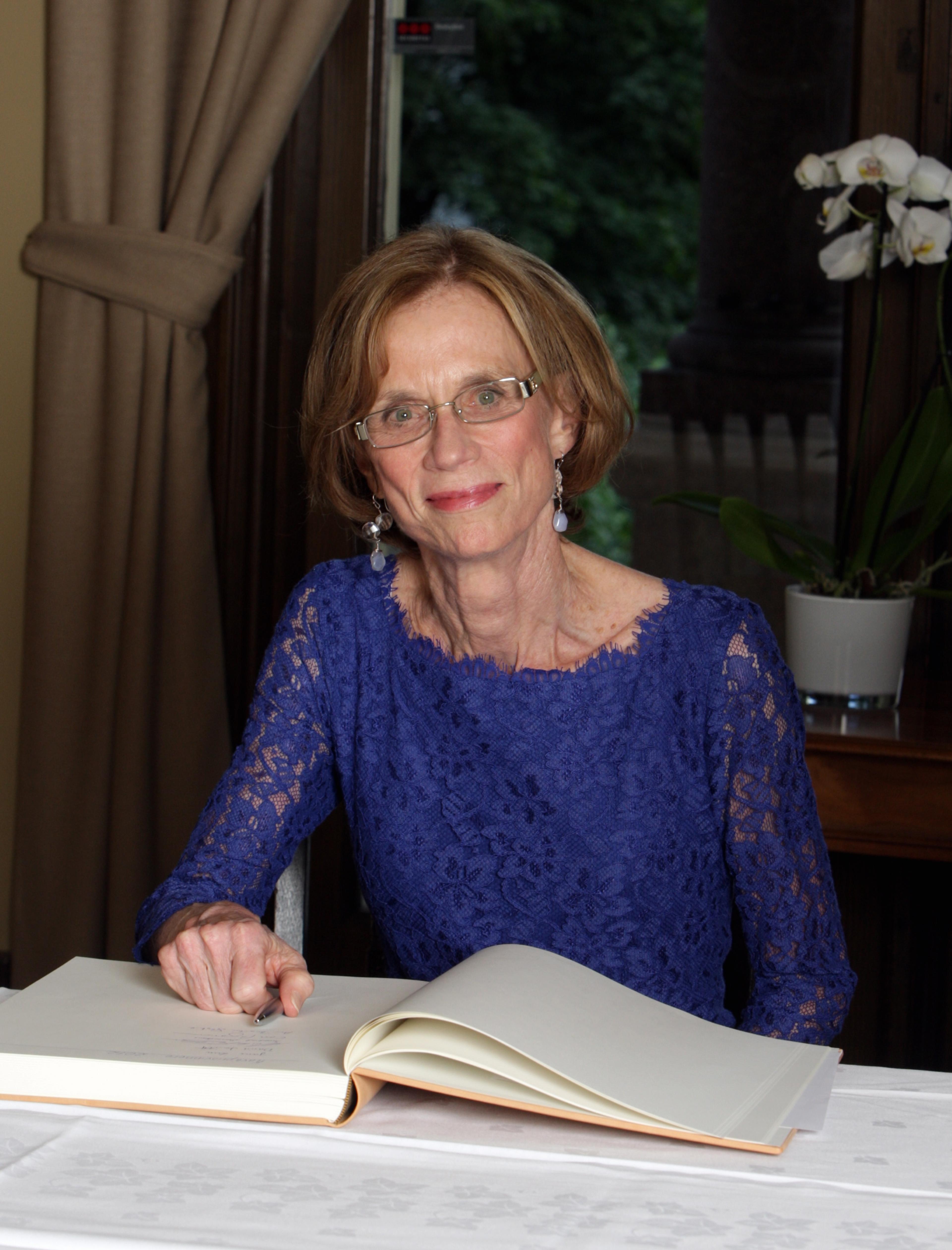
Ann Martin Graybiel signing the guest book at the Norwegian Academy of Science and Letters during the Kavli Prize week in Oslo (Photo credit: Eirik Furu Baardsen).
I then went to Harvard University and was introduced to chemistry by Eugene Rocow (famous for silicones), who included me in the first small-group freshman seminar in chemistry at Harvard, where I, totally naive about chemistry, was in the company of other students, many of whom had had chemistry labs in their basements. I decided to be a chemist! But I changed again, to major in biology. That was lucky, because I was allowed to do a research project in the laboratory of Donald Griffin, discoverer of echolocation in bats, who was fascinated by animal communication. I could hardly believe my good luck when he welcomed me to his lab and introduced me to field work in Trinidad. My first small student project was to study wiederorientierung, a form of habitual behavior in bats. Years later, I wound up studying habits by following a very different path.
Although schooled away from home, I had the chance to talk for many hours with my father during those years, He gave me the invaluable example of a person deeply inspired by the natural world and by human behavior, excited to do research, endlessly energetic, and combining his serious work with a sense of fun that was captivating. I like to think that my brother, a physician, and I split up these interests: he became a practicing physician, and I became a scientist interested in issues faced in medicine.
MIT
I received graduate training at MIT. I had the good fortune to be there during the "Teuber Era" (named for Hans-Lukas Teuber), when the MIT psychology Department was a magnet for many of the scientists pioneering the young field of neuroscience. I did my thesis work in neuroanatomy under Walle J.H. Nauta, and had a chance to do 'long shot’ experiments with Jerry Lettvin. David Hubel and Torsten Wiesel were doing their now-famous experiments at Harvard Medical School. Torsten kindly agreed to be the outside examiner for my thesis, and after graduating, I was urged to do physiology, not anatomy, by David Hubel, who had me do seven key experiments (on orientation columns) with him while Wiesel was away. I thus had the great opportunity not only to learn anatomy, but also to be exposed to physiology, to which my group and I turned many years later. It was an incredibly exciting time — neuroscience was being born as a discipline, and new techniques were beginning to hint at what might be done to learn about the brain. We knew so little! There was so much to learn!
During those years most attention turned — understandably — to the neocortex, long considered the 'highest' part of the brain functionally and evolutionarily. Hubel and Wiesel and Mountcastle discovered cortical columns; a rational ordering of cortical layers and columns became a candidate functional architecture of the neocortex. Many experiments began to chip away at questions such as how can we see? Feel textures? Sense sounds? And even, what activity goes on in the motor cortex as movements are planned? The visual system became the center of focus in much of North American neuroscience. I myself worked for some years, as a student and as a beginning MIT faculty member, to discover how the outlying cortical visual areas, and midbrain and thalamic regions, were organized. I was lucky to have David Berson as a student working with me on these problems. When we finally were able to use retrograde labeling, I jumped to look at oculomotor and visuo-oculomotor pathways.
But I was looking for a way to connect these experiments to the human brain. There was no fMRI; PET scans were new; and no tracing of connections was possible in post-mortem brain specimens (though I tried hard for a while). I came up with the idea of trying to find stains for chemicals related to neurotransmission that could be used for both experimental material and human brain material.

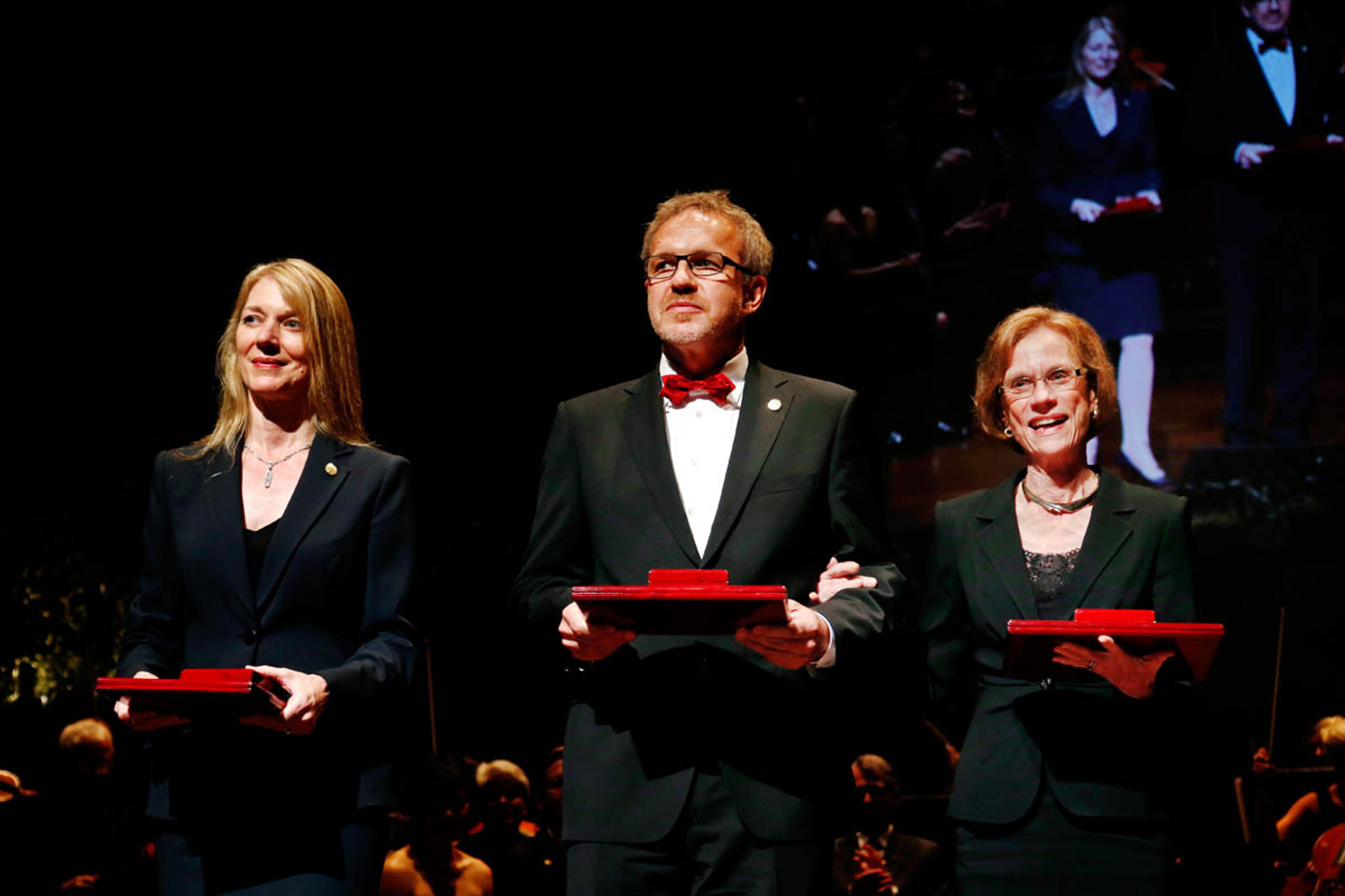
2012 Kavli Prize Neuroscience laureates at the ceremony in Oslo, from left: Cornelia Bargmann, Winfried Denk and Ann Martin Graybiel (Photo credit: Scanpix).
This strategy turned out to be useful; in animals, we were able to use an enzymatic stain related to cholinergic neurotransmission to delineate functional subdivisions in the thalamus and the superior colliculus that we had discovered by tracing pathways. We could see some of the same subdivisions in the human brain samples. But it was in looking at the striatum, a deep-lying part of the forebrain, that a quite unexpected finding emerged in experiments with Cliff Rasdale, then an MIT undergraduate, and Henry Hall, an MIT graduate working in the laboratory.
Sophisticated chemical architecture
At the time, little was known about the neuroscience of the striatum, but it was known to be the largest nucleus within the deep-forebrain system called the basal ganglia. Anatomists and others thought the striatum was a primitive brain region, because it seemed to be an undifferentiated mass of neurons lacking the beautiful layers and apical dendritic organization of the overlying neocortex. The physiology of these structures was almost unknown except for a few pioneering studies. But these structures were well known to physicians as being implicated in motor disorders, including Parkinson’s disease. The famous cholinergic-dopaminergic balance hypothesis of extrapyramidal motor control was based largely on notions about the interactions of these transmitters in the striatum.
What we saw with our cholinergic enzyme stains, first in the human brain and then in the brains of a wide range of mammalian species, was that the striatum was not primitive after all. Instead, this brain region had a sophisticated chemical architecture, whose building blocks were chemically distinct compartments hidden from view in classical stains. We named these striosomes (for striatal bodies), and found that the striosomes formed a three-dimensional labyrinth embedded in the large surrounding region, the striatal matrix.
Explorations of this striosome-matrix architecture eventually became a main focus of our lab. Soon we learned that striosomes in some ways resembled a cortical layer (for example, neurons in striosomes are born in a special time-window, like a layer of the neocortex (specifically, layer 6)). We found that during development, striosomes corresponded to the developmental 'dopamine islands' that had been discovered by Dahlstrom and Fuxe. And we, and others, found that an astonishing number of the neuropeptides, neurotransmitters and receptors, and transmitterrelated molecules followed the striosome-matrix architecture in their striatal distributions. The striosomes turned out to match the opioid receptor-rich zones observed by Solomon Snyder and his group. I was lucky in this period to work with Fu-Chin Lu and a series of extremely talented students and postdoctoral fellows, including Mary Nastuk, Marie-Francoise Chesselet, Etienne Hirsch, and Bruce Quinn and with others including Suzanne Roffler-Tarlov and Marie-Jo Besson. We concluded that this striosome-matrix architecture, which had been hidden from view in classical stains, might be a general organizing principle for the molecular anatomy of the striatum. But what did this mean functionally?

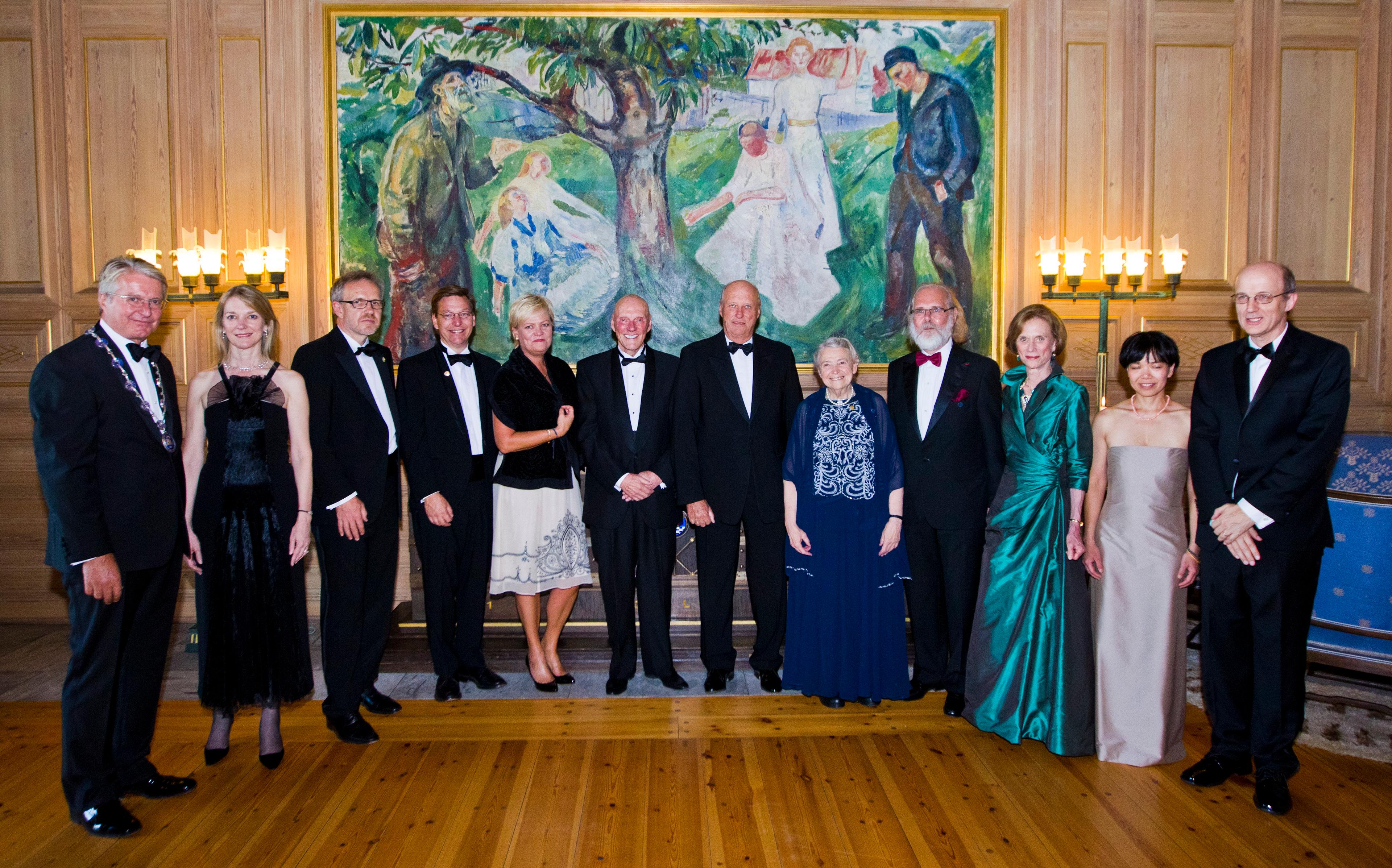
Reception in the Munch room at Oslo City Hall, from left: Mayor of Oslo Fabian Stang, Cornelia Bargmann (Kavli Prize neuroscience laureate), Winfried Denk (Kavli Prize neuroscience laureate), Michael Brown (Kavli Prize astrophysics laureate), Kristin Halvorsen (Norwegian minister of science and education), Fred Kavli, His Royal Highness King Harald, Mildred S. Dresselhaus (Kavli Prize nanoscience laureate), Nils Christian Stenseth (President of the Norwegian Academy of Science and Letters), Ann Martin Graybiel (Kavli Prize neuroscience laureate), Jane X. Luu (Kavli Prize astrophysics laureate) and David Jewitt (Kavli Prize astrophysics laureate). (Photo credit: Scanpix).
Striatum – a learning machine
A big step at this early time came when we combined the chemical stains with tracing the inputoutput connections of the striatum. It turned out that they followed this architecture also. Moreover, striosomes were preferentially related to limbic ("emotion-related") pathways, whereas the matrix was related to sensorimotor and associative regions. Most strikingly, Rafi Malach and I discovered that the huge matrix compartment was itself divided up into modules (we called them "matrisomes"), though these also could be seen in classical cell or fiber stains. Rafi was one of the two brave physiologists who came to the lab — Carl Olson was the first. Our experiments, and then those with Alice Flaherty and Hemai Parthasarathy, and with Juan Jimenez-Castillanos and Jose Gimenez-Amaya, suggested that cortical projections to the striatum are systematically remapped onto dispersed striatal modules, and that the outputs of these modules could converge in the output nuclei of the basal ganglia.
This divergence-reconvergence architecture reminded me of the mixture-of-experts computational learning architecture then being worked on by Michael Jordan and his colleagues. This could mean that the striatum might be a learning device embedded within basal ganglia circuitry. Our work on the corticostriatal remapping was happening just as Ranulfo Romo and Wolfram Schultz were discovering that the dopamine-containing neurons that project to the striatum responded to rewards, not to movements, and that these neurons acquired learning-related responses as animals were trained to do simple tasks. And soon, with Minoru Kimura and his coworker, Aosaki, we found that the neurons thought (and now known) to be the cholinergic interneurons of the striatum had responses that also changed with learning, and that if we depleted the dopamine in the striatum, these acquired responses were largely lost.
We decided to put the idea that the striatum is a learning machine to the test by adopting the ensemble recording methods just being worked out for recording in the hippocampus of freely moving rats — the tetrode recoding method of McNaughton, Matt Wilson, and others. We were lucky again. Mandar Jog, Chris Connolly, Yasuo Kubota and I had rats learn simple maze tasks, and we trained them extensively so that they eventually learned the maze runs as habits. All the while we recorded from ensembles of neurons in the sensorimotor striatum. What we found was that if we viewed the neural activity across the entire behavioral task-time, we could see a distributed pattern of plasticity that changed enormously as the animals learned: at first the ensembles of striatal neurons, on average, fired throughout the runs, but they later fired most vigorously at the beginning and end of the runs.
Learning-related experiments
This seemed to call for primate physiology, and with other brave postdoctoral fellows, including Jun Kojima, Pablo Blazquez and Naotaka Fujii, and equally brave students, including Theresa Desrochers and Joey Feingold, we developed a chronic multi-electrode system that let us do what we had been doing in the rodents. Naotaka and I found in the primate striatum and prefrontal cortex a task-bracketing pattern remarkably similar to what we had found in the rodents. It was as though action boundaries were formed when entire behaviors became well learned.
These findings suggested that one learning-related function of the striatum could be to build representations of entire repeatedly positively reinforced (advantageous) behaviors — a function analogous to the famous idea of chunking proposed in memory studies by George Miller. If so, the stream of behavior — and conceivably the stream of thought — could be organized into subunits that are manageable as parts of larger representational repertoires. We have continued this line of learning-related experiments with a series of gifted students and postdoctoral fellows (including Terra Barnes and Katy Thorn, Mark Howe, Hisham Atallah and Ledia Hernandez). Now, Kyle Smith and I are finding that we can toggle habits off and on, on-line, by optogenetic manipulations of the habit circuit.

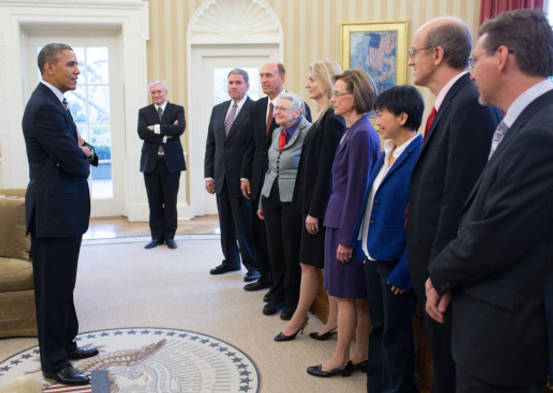
President Barack Obama greets the 2012 U.S. Kavli Prize Laureates in the Oval Office, March 28, 2013. Clockwise from the President: Wegger Chr. Strommen, Norwegian Ambassador to the United States; Rockell N. Hankin, Vice-Chairman of The Kavli Foundation; Robert W. Conn, President of The Kavli Foundation; Kavli Laureates Mildred S. Dresselhaus, Cornelia Isabella Bargmann, Ann M. Graybiel, Jane X. Luu, David C. Jewitt and Michael E. Brown. (Official White House Photo by Pete Souza)
Repetitive behavior
At the same time, following on work that Richard Courtemanche and I did in primates, we have come to focus also on the highly dynamic patterns of oscillatory activity visible in local field potentials that occur when animals learn and perform habits. Bill DeCoteau, Katy Thorn, Joey Feingold, Mark Howe and Dan Gibson have all been important in this work.
A powerful way to induce a habit, including an unwanted habit, is to use addictive drugs. I decided to give rats amphetamine and to use the then very new technique of staining for the de novo expression of early response genes. The result was amazing. We could see striosomes for the first time in a functional assay! Luckily, Rosario Moratalla joined the lab, and then Sabina Berretta, Juan Canales, Christine Capper-Loupe, Bulant Elibol, and Essen Saka. We and our coworkers found that "striosome predominance" of the gene induction was closely correlated with the degree to which the animals expressed drug-induced stereotypies.
This was intriguing: could striosomes have something to do with the compulsion to repeat a chosen behavior? A critical piece of the puzzle came when Frank Eblen and I found that the far-anterior (pregenual) anterior cingulate cortex and the posterior orbitofrontal cortex preferentially project to striosomes. These were, as others had found, paired cortical sites in the "OCD circuit" related to mood and to compulsive repetitive behaviors in humans: in brain scans, they appeared abnormal in individuals with obsessive compulsive disorder or suffering from addictive states. And we and others had suggestive evidence that striosomes might be the origin of pathways controlling the dopamine-containing neurons in the substantia nigra. This striosome-dopamine circuit might be critical for behavioral control.
We clearly needed causal evidence. I went back to school to learn enough molecular biology to allow us to try to clone striatum-enriched genes, and with Hiroaki Kawasaki and David Housman and our coworkers, we discovered then-novel families of genes that target the ras-superfamily. Two of these turned out to be differentially expressed in striosomes (CalDAG-GEFII) and matrix (CalDAG-GEFI). Fortunately, Jill Crittenden came to the lab and engineered mouse knockouts of the genes, and we have found that knockouts lacking the matrix-enriched gene have exaggerated repetitive behaviors and are overly focused. Now Eric Burguiere and I are tackling this issue with optogenetics, and finding that manipulations of related circuits can stop compulsive behavior. With Richard Faull and his New Zealand group, we found that Huntington's disease patients with mood disturbances as early primary symptoms — including repetitive behaviors — have differential loss of striosomes compared to patients with primary movement problems as early symptoms.
And critically, Ken-ichi Amemori and I formulated the hypothesis that cost-benefit decision-making might be an essential function of these striatal compartments. We now have evidence that such decisions can be strongly biased by microstimulation of what we think is striosome-projecting anterior cingulate cortex, and Alexander Friedman and Leif Gibb have intriguing evidence on this point.
Continual exploration of the brain
These sketches of our research make what to me is a very important point: We started out with a set of findings in basic science, by celebrating (and we did!) the excitement of discovering new features of the brain's organization and function. But by keeping on with these lines of work, we now are naturally approaching the point at which we can search for ways that this research could help people. I have had the privilege of working closely with physician-scientists, and through these collaborations, with Richard Faull (for Huntington's disease), William Langston (for Parkinson's disease), and Ryuji Kaji and Satoshi Goto (for dystonias), we finally can begin to link striosomematrix architecture to patterns of neurodegeneration in conditions that afflict humans. We have hints, strong hints, for a relation to certain neuropsychiatric disorders as well, but these must be explored more. For us, there is no conflict between pure research and translational research — they blend inextricably in work on the basal ganglia. We are determined to continue to explore the amazing functional organization of the brain and, if we can, to uncover mechanisms of action of cortico-basal ganglia circuits that will yield critical insights into these disorders and the potential for their treatment.
This is an autobiography full of science, and maybe not so full of personal detail. Our lab has run almost as a family, and I am grateful to have the chance to recall such a wonderful group of students, fellows and collaborators. I have been fortunate to work for many years with Diane Major, Henry Hall, Hu Dan, Pat Harlan and Margo Cantor, definitely family. And I have been helped by many people who have encouraged us when the going was tough. Outside of work, I take special delight in spending time in the natural world, in music, in friendships, and in physically active engagement ranging from heavy outside work to sports including tennis, swimming, and cycling in beautiful places. I have, encouraged by the lab work, an ever-increasing interest in rituals and in our behaviors as humans adapting to the world as we know it and guess it to be. Most important, I had the good fortune to have met and become the bride of Jim Lackner, whom I met at MIT during graduate school under the encouragement of Hans-Lukas Teuber. Marvelously talented and inventive, and with both undergraduate and graduate degrees from MIT and a fascinating career working on human spatial orientation, my husband manages to make life very interesting indeed.